

Dynamics and mechanisms of posterior axis elongation in the vertebrate embryo
- PMID: 30283977
- PMCID: PMC11105343
- DOI: 10.1007/s00018-018-2927-4
Dynamics and mechanisms of posterior axis elongation in the vertebrate embryo
Abstract
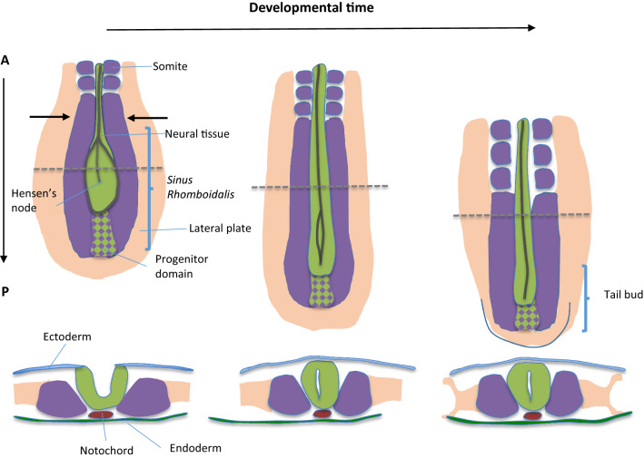
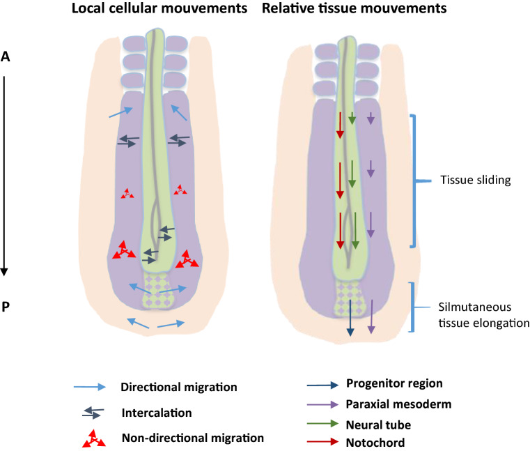
During development, the vertebrate embryo undergoes significant morphological changes which lead to its future body form and functioning organs. One of these noticeable changes is the extension of the body shape along the antero-posterior (A-P) axis. This A-P extension, while taking place in multiple embryonic tissues of the vertebrate body, involves the same basic cellular behaviors: cell proliferation, cell migration (of new progenitors from a posterior stem zone), and cell rearrangements. However, the nature and the relative contribution of these different cellular behaviors to A-P extension appear to vary depending upon the tissue in which they take place and on the stage of embryonic development. By focusing on what is known in the neural and mesodermal tissues of the bird embryo, I review the influences of cellular behaviors in posterior tissue extension. In this context, I discuss how changes in distinct cell behaviors can be coordinated at the tissue level (and between tissues) to synergize, build, and elongate the posterior part of the embryonic body. This multi-tissue framework does not only concern axis elongation, as it could also be generalized to morphogenesis of any developing organs.
Keywords: Axis elongation; Bird embryo; Live imaging; Morphogenesis; Multi-tissue; PSM; Proliferation; Tissue deformations.
Figures
Similar articles
-
Mechanics of Anteroposterior Axis Formation in Vertebrates.Annu Rev Cell Dev Biol. 2019 Oct 6;35:259-283. doi: 10.1146/annurev-cellbio-100818-125436. Epub 2019 Aug 14. Annu Rev Cell Dev Biol. 2019. PMID: 31412208 Free PMC article. Review.
-
Multi-scale quantification of tissue behavior during amniote embryo axis elongation.Development. 2017 Dec 1;144(23):4462-4472. doi: 10.1242/dev.150557. Epub 2017 Aug 23. Development. 2017. PMID: 28835474
-
[A non directional cell migration gradient in the presomitic mesoderm contributes to axis elongation in chicken embryos].Biol Aujourdhui. 2011;205(2):95-103. doi: 10.1051/jbio/2011014. Epub 2011 Aug 11. Biol Aujourdhui. 2011. PMID: 21831340 French.
-
Differential proliferation regulates multi-tissue morphogenesis during embryonic axial extension: integrating viscous modeling and experimental approaches.Development. 2024 Jul 1;151(13):dev202836. doi: 10.1242/dev.202836. Epub 2024 Jul 4. Development. 2024. PMID: 38856082
-
Formation and segmentation of the vertebrate body axis.Annu Rev Cell Dev Biol. 2013;29:1-26. doi: 10.1146/annurev-cellbio-101011-155703. Epub 2013 Jun 26. Annu Rev Cell Dev Biol. 2013. PMID: 23808844 Review.
Cited by
-
Extracellular volume expansion drives vertebrate axis elongation.Curr Biol. 2025 Feb 24;35(4):843-853.e6. doi: 10.1016/j.cub.2024.12.051. Epub 2025 Jan 28. Curr Biol. 2025. PMID: 39879975 Free PMC article.
-
Mechanics of Anteroposterior Axis Formation in Vertebrates.Annu Rev Cell Dev Biol. 2019 Oct 6;35:259-283. doi: 10.1146/annurev-cellbio-100818-125436. Epub 2019 Aug 14. Annu Rev Cell Dev Biol. 2019. PMID: 31412208 Free PMC article. Review.
-
Programming the elongation of mammalian cell aggregates with synthetic gene circuits.bioRxiv [Preprint]. 2024 Dec 11:2024.12.11.627621. doi: 10.1101/2024.12.11.627621. bioRxiv. 2024. PMID: 39713354 Free PMC article. Preprint.
-
Spinal cord elongation enables proportional regulation of the zebrafish posterior body.Development. 2025 Jan 1;152(1):dev204438. doi: 10.1242/dev.204438. Epub 2025 Jan 9. Development. 2025. PMID: 39745249 Free PMC article.
References
-
- Keller RE, Danilchik M, Gimlich R, Shih J. The function and mechanism of convergent extension during gastrulation of Xenopus laevis. J Embryol Exp Morphol. 1985;89(Suppl):185–209. - PubMed
Publication types
MeSH terms
LinkOut - more resources
Full Text Sources
Miscellaneous
