. 2018 Dec;136(6):939-953.
doi: 10.1007/s00401-018-1915-y. Epub 2018 Oct 3.

Mutant superoxide dismutase aggregates from human spinal cord transmit amyotrophic lateral sclerosis

Affiliations

Mutant superoxide dismutase aggregates from human spinal cord transmit amyotrophic lateral sclerosis

Elaheh Ekhtiari Bidhendi et al. Acta Neuropathol. 2018 Dec.

Abstract

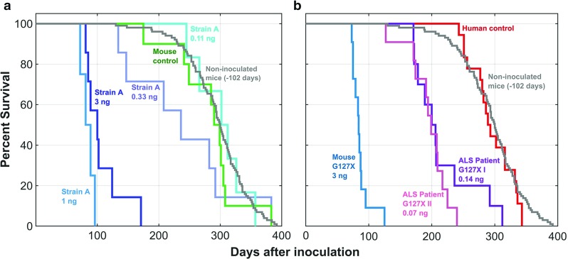
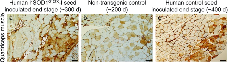
Motor neurons containing aggregates of superoxide dismutase 1 (SOD1) are hallmarks of amyotrophic lateral sclerosis (ALS) caused by mutations in the gene encoding SOD1. We have previously reported that two strains of mutant human (h) SOD1 aggregates (denoted A and B) can arise in hSOD1-transgenic models for ALS and that inoculation of such aggregates into the lumbar spinal cord of mice results in rostrally spreading, templated hSOD1 aggregation and premature fatal ALS-like disease. Here, we explored whether mutant hSOD1 aggregates with prion-like properties also exist in human ALS. Aggregate seeds were prepared from spinal cords from an ALS patient carrying the hSOD1G127Gfs*7 truncation mutation and from mice transgenic for the same mutation. To separate from mono-, di- or any oligomeric hSOD1 species, the seed preparation protocol included ultracentrifugation through a density cushion. The core structure of hSOD1G127Gfs*7 aggregates present in mice was strain A-like. Inoculation of the patient- or mouse-derived seeds into lumbar spinal cord of adult hSOD1-expressing mice induced strain A aggregation propagating along the neuraxis and premature fatal ALS-like disease (p < 0.0001). Inoculation of human or murine control seeds had no effect. The potencies of the ALS patient-derived seed preparations were high and disease was initiated in the transgenic mice by levels of hSOD1G127Gfs*7 aggregates much lower than those found in the motor system of patients carrying the mutation. The results suggest that prion-like growth and spread of hSOD1 aggregation could be the primary pathogenic mechanism, not only in hSOD1 transgenic rodent models, but also in human ALS.

Keywords: Aggregation; Motor neuron disease; Prion-like; Propagation; Superoxide dismutase.


Conflict of interest statement

None of the authors report any conflicts of interest.

Figures

Fig. 1
Fig. 1
Binary epitope-mapping patterns of hSOD1 aggregates in spinal cords from end-stage non-inoculated and inoculated transgenic mice. Results for antibodies raised against longer SOD1 peptides used in the original protocol are labeled blue, and data for the additional antibodies raised against shorter peptides are labeled red. The aggregates were analyzed in 5 end-stage mice in all groups and the standard deviations and means are shown. To facilitate appreciation of patterns, all results were normalized against the staining intensity with the 57-72 Ra-Ab (= 100%). a and b Non-inoculated hSOD1G85R and hSOD1G127X Tg mice, respectively. cf hSOD1G85R Tg mice inoculated with indicated seeds
Fig. 2
Fig. 2
Inoculation of hSOD1 aggregate-containing seeds caused premature fatal motor neuron disease. a Inoculation of different dilutions of a hSOD1G85R strain A seed compared with a non-transgenic control mouse seed. b Inoculation of a human and mouse-derived hSOD1G127X seeds compared with a human control seed. As references both figures contain survival data for non-inoculated hSOD1G85R Tg mice (n = 101) subtracted with the mean age (102 days) at which the inoculations took place. In a the post-inoculation survival lengths and significances for differences versus the mouse control were for the 3, 1, and 0.33 ng inoculations 107 ± 31 days, p < 0.0001; 85 ± 10 days, p < 0.002; and 301 ± 41, p < 0.14 (ns), respectively. The 0.33 ng data were significantly different from those of the non-inoculated mice (p < 0.04). For analysis of data in b. see Table 1
Fig. 3
Fig. 3
Fiber-type grouping in end-stage mice. a, c Micrographs of quadriceps muscle from end-stage hSOD1G85R Tg mice inoculated with the human hSOD1G127X–I or control seeds and b a 200-day-old C57BL/6 control mouse. The muscles were stained with an antibody against slow myosin and the micrographs from the end-stage mice shows fiber type grouping indicating denervation-induced atrophy. Scale bars = 50 µm
Fig. 4
Fig. 4
Content of strain A aggregates in whole spinal cords of inoculated and non-inoculated hSOD1G85R Tg mice analyzed with binary epitope mapping. Triangles (triangle), crosses (cross) and circles (unfilled circle) indicate non-symptomatic, symptomatic and end-stage mice, respectively. Blue, violet, green, and red symbols indicate hSOD1G85R Tg mice inoculated with the mouse hSOD1G127X (blue unfilled triangle, blue unfilled cross, blue unfilled circle), human hSOD1G127X I (purple unfilled triangle, purple unfilled cross, purple unfilled circle), mouse control (green unfilled triangle, green unfilled circle) and human control (red unfilled triangle, red unfilled circle) seeds, respectively. Gray indicates non-inoculated mice (gray unfilled triangle, gray unfilled circle) (lifespans minus 102 days, the mean age at which the inoculations took place). The shaded area indicates blank reactions
Fig. 5
Fig. 5
Distribution of strain A hSOD1 aggregation along the neuraxis in individual end-stage Tg mice. The aggregates were analyzed with binary epitope mapping using the 57–72 Ra-Ab. The levels in the segments were normalized against the levels in the lumbar spinal cord. Note the change in scale for brain and cerebellum. The results for individual mice are presented in different colors to improve distinction. a, d Results for hSOD1G85R Tg mice inoculated with indicated seeds. In a, b, and d only the left (inoculation side, closed circles) or right halves (open circles) of the neuraxes were analyzed. In b, results for the two human hSOD1G127X-I seed-inoculated mice with lifespans equal to or longer than the mean survivals of the human control-inoculated and the non-inoculated mice are labeled (cross) (c.f. Fig. 2b). e, f Results for non-inoculated hSOD1G127X and hSOD1G85R Tg mice
Fig. 6
Fig. 6
Histopathology of end-stage inoculated and asymptomatic non-inoculated hSODG85R Tg mice. a–l Sections were stained with the 131–153 Mo-Ab. For the non-inoculated 300-day-old control mice (middle column), a typical picture for the five mice showing sparse labeling is shown. In seven, no labeling was found and in one, the staining was comparable to that of end-stage mice. Note that while non-inoculated 300-day-old hSOD1G85R Tg mice almost entirely show thread-like immunoreactivity (b, e, h, and k), the end-stage inoculated mice show widespread granular reactivity in motor neurons somas as well as neuropil threads (a, c, d, f, g, i, j, l). m–o Sections were stained with the 131–153 Ch-Ab (green) and an antibody against GFAP (red). Yellow arrows indicate colocalization of misfolded SOD1 and GFAP. Note the large difference between hSOD1G127X-I seed-inoculated (m) and age-matched control animals (n). pr sections from end stage non-inoculated hSOD1G85R Tg mice stained with the 131–153 Mo-Ab. Scale bars = 10 µm

References

    1. Andersen PM, Al-Chalabi A. Clinical genetics of amyotrophic lateral sclerosis: what do we really know? Nat Rev Neurol. 2011;7:603–615. doi: 10.1038/nrneurol.2011.150. - DOI - PubMed
    1. Andersen PM, Nilsson P, Keranen ML, Forsgren L, Hagglund J, Karlsborg M, Ronnevi LO, Gredal O, Marklund SL. Phenotypic heterogeneity in motor neuron disease patients with CuZn-superoxide dismutase mutations in Scandinavia. Brain. 1997;120(Pt 10):1723–1737. doi: 10.1093/brain/120.10.1723. - DOI - PubMed
    1. Ayers JI, Diamond J, Sari A, Fromholt S, Galaleldeen A, Ostrow LW, Glass JD, Hart PJ, Borchelt DR. Distinct conformers of transmissible misfolded SOD1 distinguish human SOD1-FALS from other forms of familial and sporadic ALS. Acta Neuropathol. 2016;132:827–840. doi: 10.1007/s00401-016-1623-4. - DOI - PMC - PubMed
    1. Ayers JI, Fromholt S, Koch M, DeBosier A, McMahon B, Xu G, Borchelt DR. Experimental transmissibility of mutant SOD1 motor neuron disease. Acta Neuropathol. 2014;128:791–803. doi: 10.1007/s00401-014-1342-7. - DOI - PubMed
    1. Ayers JI, Fromholt SE, O’Neal VM, Diamond JH, Borchelt DR. Prion-like propagation of mutant SOD1 misfolding and motor neuron disease spread along neuroanatomical pathways. Acta Neuropathol. 2016;131:103–114. doi: 10.1007/s00401-015-1514-0. - DOI - PMC - PubMed

Publication types

Substances