

Altered Amniotic Fluid Levels of Hyaluronic Acid in Fetal Rats with Myelomeningocele: Understanding Spinal Cord Injury
- PMID: 30284959
- PMCID: PMC7718849
- DOI: 10.1089/neu.2018.5894
Altered Amniotic Fluid Levels of Hyaluronic Acid in Fetal Rats with Myelomeningocele: Understanding Spinal Cord Injury
Abstract
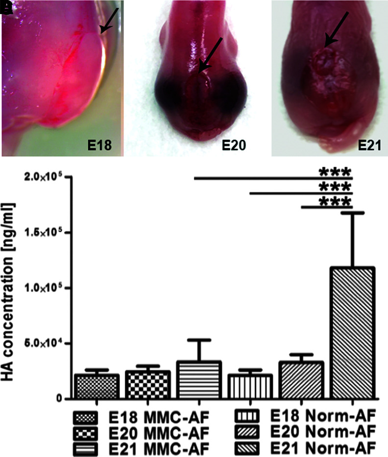
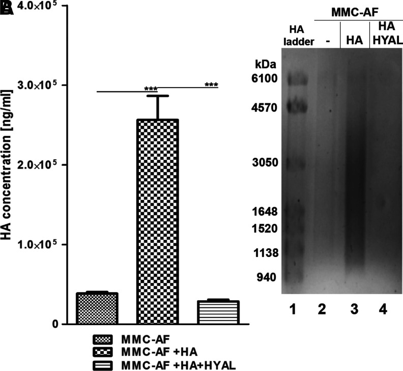
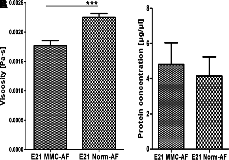
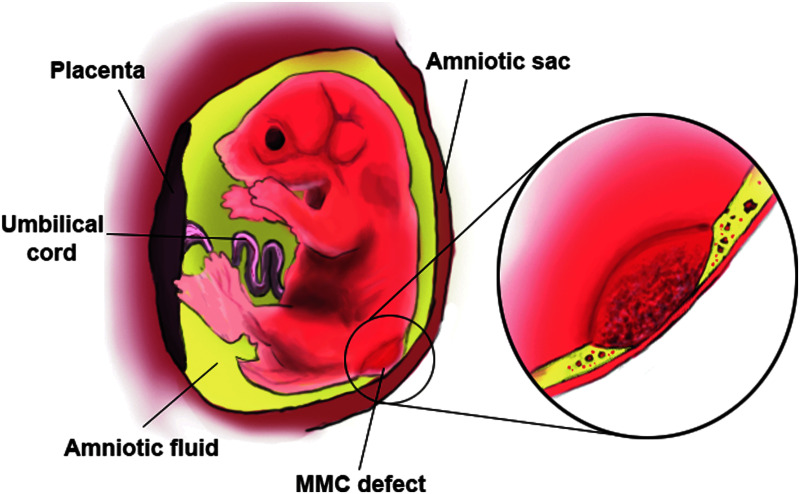
Myelomeningocele (MMC) is a devastating congenital neural tube defect that results in the exposure of spinal cord to the intrauterine environment, leading to secondary spinal cord injury and severe impairment. Although the mechanisms underlying the secondary pathogenesis are clinically relevant, the exact cause of in utero-acquired spinal cord damage remains unclear. The objective of this study was to determine whether the hyaluronic acid (HA) concentration in amniotic fluid (AF) in the retinoic acid-induced model of MMC is different from that in normal controls and whether these differences could have an impact on the viscosity of AF. Our data shows that the concentration of HA in AF samples from fetuses with MMC (MMC-AF) and normal control samples (Norm-AF) were not significantly different at embryonic day 18 (E18) and E20. Thereafter, the HA concentration significantly increased in Norm-AF but not in MMC-AF. Compared with Norm-AF, the concentration of HA in MMC-AF and the viscosity of MMC-AF were significantly lower at E21. Agarose gel electrophoresis confirmed a significant reduction in the HA level of MMC-AF compared with Norm-AF at E21. No HA-degrading activity was detected in MMC-AF. In summary, we identified a deficiency in the AF level of HA and the viscosity of AF in fetal rats with MMC. These data are discussed in relation to a potential role the reduction in the AF viscosity due to the low level of HA may play in the exacerbating effects of mechanical trauma on spinal cord damage at the MMC lesion site.
Keywords: fetal rats; hyaluronic acid; myelomeningocele; neural tube defects; spinal cord injury.
Conflict of interest statement
No competing financial interests exist.
Figures

Similar articles
-
Amylase concentration and activity in the amniotic fluid of fetal rats with retinoic acid induced myelomeningocele.J Matern Fetal Neonatal Med. 2022 Jan;35(1):147-154. doi: 10.1080/14767058.2020.1713082. Epub 2020 Jan 16. J Matern Fetal Neonatal Med. 2022. PMID: 31910702
-
Amniotic fluid levels of phospholipase A2 in fetal rats with retinoic acid induced myelomeningocele: the potential "second hit" in neurologic damage.J Matern Fetal Neonatal Med. 2016 Sep;29(18):3003-8. doi: 10.3109/14767058.2015.1112373. Epub 2015 Nov 23. J Matern Fetal Neonatal Med. 2016. PMID: 26513600
-
Clusters of amniotic fluid cells and their associated early neuroepithelial markers in experimental myelomeningocele: Correlation with astrogliosis.PLoS One. 2017 Mar 30;12(3):e0174625. doi: 10.1371/journal.pone.0174625. eCollection 2017. PLoS One. 2017. PMID: 28358903 Free PMC article.
-
Role of Amniotic Fluid Toxicity in the Pathophysiology of Myelomeningocele: A Narrative Literature Review.Prenat Diagn. 2024 Nov;44(12):1530-1535. doi: 10.1002/pd.6681. Epub 2024 Oct 6. Prenat Diagn. 2024. PMID: 39370541 Review.
-
Preclinical stem cell therapy in fetuses with myelomeningocele: A systematic review and meta-analysis.Prenat Diagn. 2021 Feb;41(3):283-300. doi: 10.1002/pd.5887. Epub 2021 Jan 11. Prenat Diagn. 2021. PMID: 33427329 Free PMC article.
Cited by
-
State of the art in translating experimental myelomeningocele research to the bedside.Childs Nerv Syst. 2021 Sep;37(9):2769-2785. doi: 10.1007/s00381-021-05299-1. Epub 2021 Jul 31. Childs Nerv Syst. 2021. PMID: 34333685 Review.
-
Spinal Cord Organoids from Human Amniotic Fluid iPSC Recapitulate the Diversity of Cell Phenotypes During Fetal Neural Tube Morphogenesis.Mol Neurobiol. 2025 Aug;62(8):10954-10969. doi: 10.1007/s12035-025-04944-z. Epub 2025 Apr 20. Mol Neurobiol. 2025. PMID: 40254702
-
Identification of Neurocan and Phosphacan as Early Biomarkers for Open Neural Tube Defects.Cells. 2023 Apr 4;12(7):1084. doi: 10.3390/cells12071084. Cells. 2023. PMID: 37048157 Free PMC article.
-
Spinal Cord Injury in Myelomeningocele: Prospects for Therapy.Front Cell Neurosci. 2020 Jun 30;14:201. doi: 10.3389/fncel.2020.00201. eCollection 2020. Front Cell Neurosci. 2020. PMID: 32714152 Free PMC article. Review.
-
Research progress on ultrasound and molecular markers for prenatal diagnosis of neural tube defects.Heliyon. 2024 Aug 13;10(16):e36060. doi: 10.1016/j.heliyon.2024.e36060. eCollection 2024 Aug 30. Heliyon. 2024. PMID: 39247260 Free PMC article. Review.
References
-
- Meuli M. and Moehrlen U. (2013). Fetal surgery for myelomeningocele: a critical appraisal. Eur. J. Pediatr. Surg. 23, 103–109 - PubMed
-
- Parker S.E., Mai C.T., Canfield M.A., Rickard R., Wang Y., Meyer R.E., Anderson P., Mason C.A., Collins J.S., Kirby R.S., and Correa A; National Birth Defects Prevention Network. (2010). Updated National Birth Prevalence estimates for selected birth defects in the United States, 2004–2006. Birth Defects Res. A Clin Mol. Teratol. 88, 1008–1016 - PubMed
-
- Kaufman B.A. (2004). Neural tube defects. Pediatr. Clin. North Am. 51, 389–419 - PubMed
-
- Dias M.S. and McLone D.G. (1993). Hydrocephalus in the child with dysraphism. Neurosurg. Clin. N. Am. 4, 715–726 - PubMed
-
- Hunt G.M. (1990). Open spina bifida: outcome for a complete cohort treated unselectively and followed into adulthood. Dev. Med. Child Neurol. 32, 108–118 - PubMed
Publication types
MeSH terms
Substances
Grants and funding
LinkOut - more resources
Full Text Sources
Other Literature Sources
