. 2018 Dec 13;122(49):11363-11372.
doi: 10.1021/acs.jpcb.8b07262. Epub 2018 Oct 17.

How the Conformations of an Internal Junction Contribute to Fold an RNA Domain

Affiliations

How the Conformations of an Internal Junction Contribute to Fold an RNA Domain

Yen-Lin Chen et al. J Phys Chem B. .

Abstract

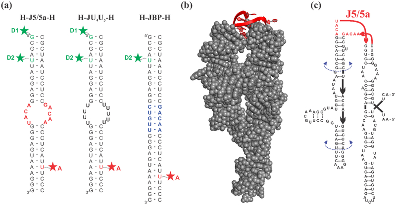
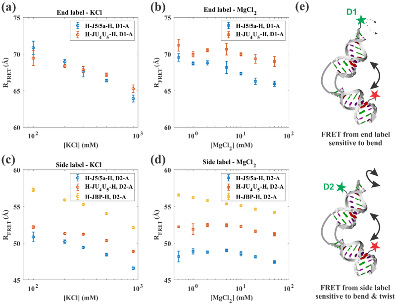
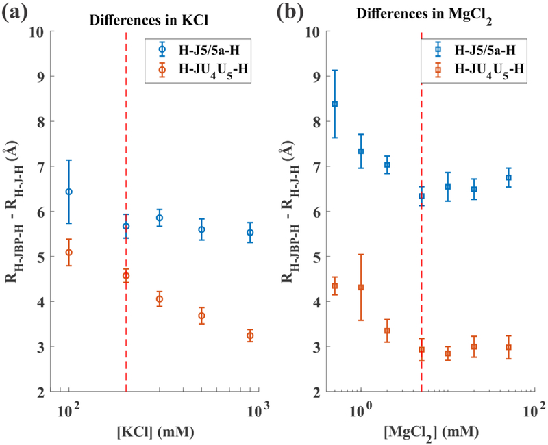
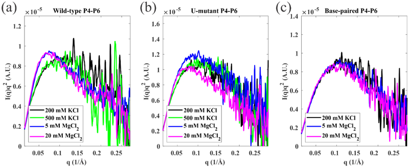
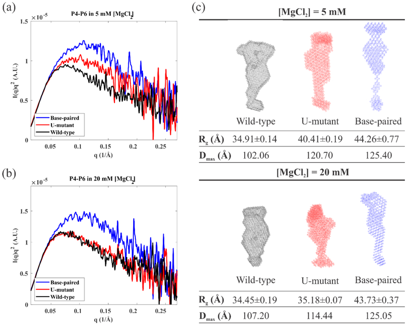
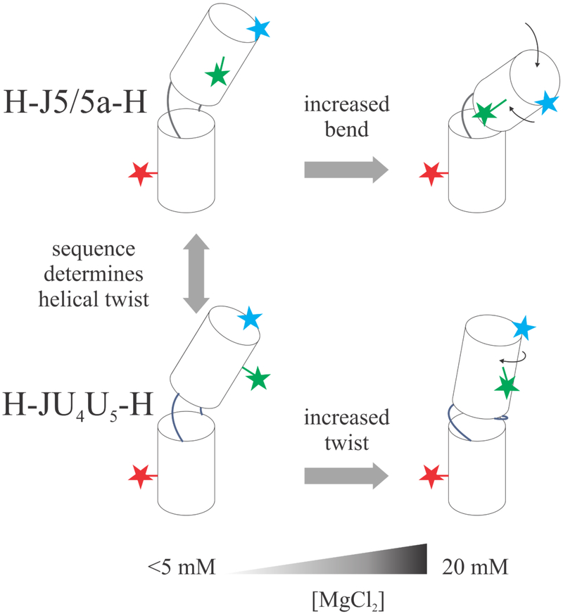
Like proteins, some RNAs fold to compact structures. We can model functional RNAs as a series of short, rigid, base-paired elements, connected by non-base-paired nucleotides that serve as junctions. These connecting regions bend and twist, facilitating the formation of tertiary contacts that stabilize compact states. Here, we explore the roles of salt and junction sequence in determining the structures of a ubiquitous connector: an asymmetric internal loop. We focus on the J5/5a junction from the widely studied P4-P6 domain of the Tetrahymena ribozyme. Following the addition of magnesium ions to fold P4-P6, this junction bends dramatically, bringing the two halves of the RNA domain together for tertiary contact engagement. Using single-molecule fluorescence resonance energy transfer (smFRET), we examine the role of sequence and salt on model RNA constructs that contain these junction regions. We explore the wild-type J5/5a junction as well as two sequence variants. These junctions display distinct, salt-dependent conformations. Small-angle X-ray scattering (SAXS) measurements verify that these effects persist in the full-length P4-P6 domain. These measurements underscore the importance of junction sequence and interactions with ions in facilitating RNA folding.


Figures

Figure 1:
Figure 1:
(a) Schematic of three RNA constructs. Constructs consist of two RNA oligonucleotides which are individually labeled with a fluorophore and annealed together. The resulting construct forms either two short duplexes connected by an asymmetric internal loop, or a fully base paired control. Green and red stars represent the dye label positions, which are achieved from either 5’ C12-amino modifier (D1) or an amino-modified dT replacing the uracil nucleotide in the sequence (D2 and A). The constructs are named according to the nature of the junction at the center. H-J5/5a-H: the junction has the native J5/5a sequence found in P4-P6. H-JU4U5-H: both junction strands are replaced by uracil nucleotides. H-JBP-H: the junction contains 5 Watson Crick base pairs. (b) The crystal structure of the full-length wild-type P4-P6 RNA (PDB: 1GID) with the J5/5a junction shown in red. (c) The sequence of the wild-type P4-P6 RNA. The U-mutant P4-P6 replaces the J5/5a junction sequence with 9 uracil nucleotides. The base-paired P4-P6 replaces “ACAU” with “CUGUU” forming 5 canonical base pairs within the junction region.
Figure 2:
Figure 2:
Measured effects on RFRET as a function of KCl and MgCl2 concentration for the D1-A (end-label) dye pair ((a) and (b)) and the D2-A (side-label) dye pair ((c) and (d)). Data for all three constructs are shown: H-J5/5a-H (blue), H-JU4U5-H (red) and H-JBP-H (orange). The error bars represent the combination of the 95% confidence interval of the fit parameter and the standard deviations of at least two independent measurements. Part (e) shows a cartoon representing the RNA junction constructs and the associated label positions. The D1 label is closer to the helical axis and is more sensitive to overall bend angle, while the D2 label is off-axis and sensitive to both bending and relative twisting.
Figure 3:
Figure 3:
The difference between RFRET for the base paired (H-JBP-H) control and H-J-H in (a) KCl and (b) MgCl2, where J represents either J5/5a or JU4U5. Dashed red lines mark a salt concentration above which one of the difference values remains constant. Note the RH –JBP – HRHJU4U5H does not approach a constant value at high concentration of KCl. Above 5 mM MgCl2 the H-JU4U5-H construct is 3 Å shorter than the base paired construct. This value is close to the height contributed by one base pair (2.8 Å) to an A-form RNA duplex.
Figure 4:
Figure 4:
Kratky plots of SAXS data reporting the salt dependence of (a) wild-type P4-P6 (b) U-mutant P4-P6 and (c) base-paired P4-P6. These plots show a more pronounced peak when the molecule is in a more compact state. The wild-type P4-P6 shows two states, unfolded (black, green, showing no peak) in KCl and folded (blue, magenta, peak appears) in MgCl2 respectively. The U-mutant P4-P6 appears unfolded in KCl, partially folded in 5 mM MgCl2 and fully folded in 20 mM MgCl2. By design the base-paired P4-P6 remains unfolded across all the conditions.
Figure 5:
Figure 5:
Comparison between the three different P4-P6 sequences in (a) 5mM MgCl2 and (b) 20mM MgCl2. (c) The reconstructions of the SAXS profiles in 5mM and 20mM MgCl2 (described in Material and Method section), color coded by the same scheme, assuming a homogeneous population. Global shape parameters are also reported.
Figure 6:
Figure 6:
Schematic representing salt concentration and sequence effects of RNA junction conformations. At low salt, the H-J5/5a-H and H-JU4U5-H have different helical twists (shown by the side label RFRET data), but similar bend angles (shown by the end label RFRET data). As [MgCl2] increases, the difference between sequences also increases, demonstrating an interplay between sequence and salt dependence.

References

    1. Sharp PA. Essay The Centrality of RNA. 2009:577–580. doi:10.1016/j.cell.2009.02.007. - DOI - PubMed
    1. Morris KV, Mattick JS. The rise of regulatory RNA. Nat Publ Gr. 2014;15(6):423–437. doi: 10.1038/nrg3722. - DOI - PMC - PubMed
    1. Cech TR, Steitz JA. Review The Noncoding RNA Revolution — Trashing Old Rules to Forge New Ones. Cell. 2014;157(1):77–94. doi:10.1016/j.cell.2014.03.008. - DOI - PubMed
    1. Schroeder R, Barta A, Semrad K. Strategies for RNA folding and assembly. Nat Rev Mol Cell Biol. 2004;5(11):908–919. doi:10.1038/nrm1497. - DOI - PubMed
    1. Al-Hashimi HM, Walter NG. RNA dynamics: it is about time. Curr Opin Struct Biol. 2008;18(3):321–329. doi:10.1016/j.sbi.2008.04.004. - DOI - PMC - PubMed

Publication types