. 2018 Nov 2;17(11):3959-3975.
doi: 10.1021/acs.jproteome.8b00618. Epub 2018 Oct 4.

Proteomic Investigation of Murine Neuronal α7-Nicotinic Acetylcholine Receptor Interacting Proteins

Affiliations

Proteomic Investigation of Murine Neuronal α7-Nicotinic Acetylcholine Receptor Interacting Proteins

Matthew J Mulcahy et al. J Proteome Res. .

Abstract

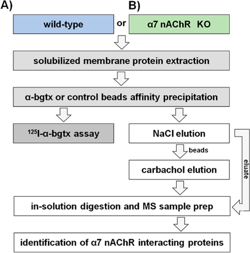
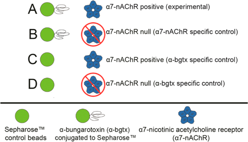
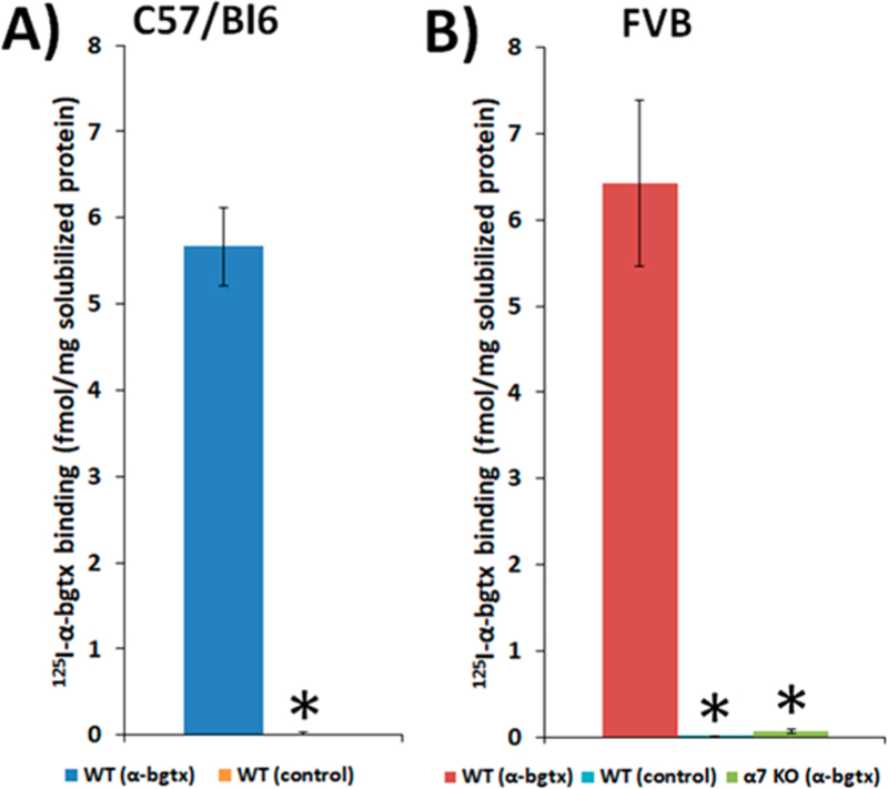
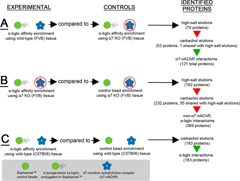
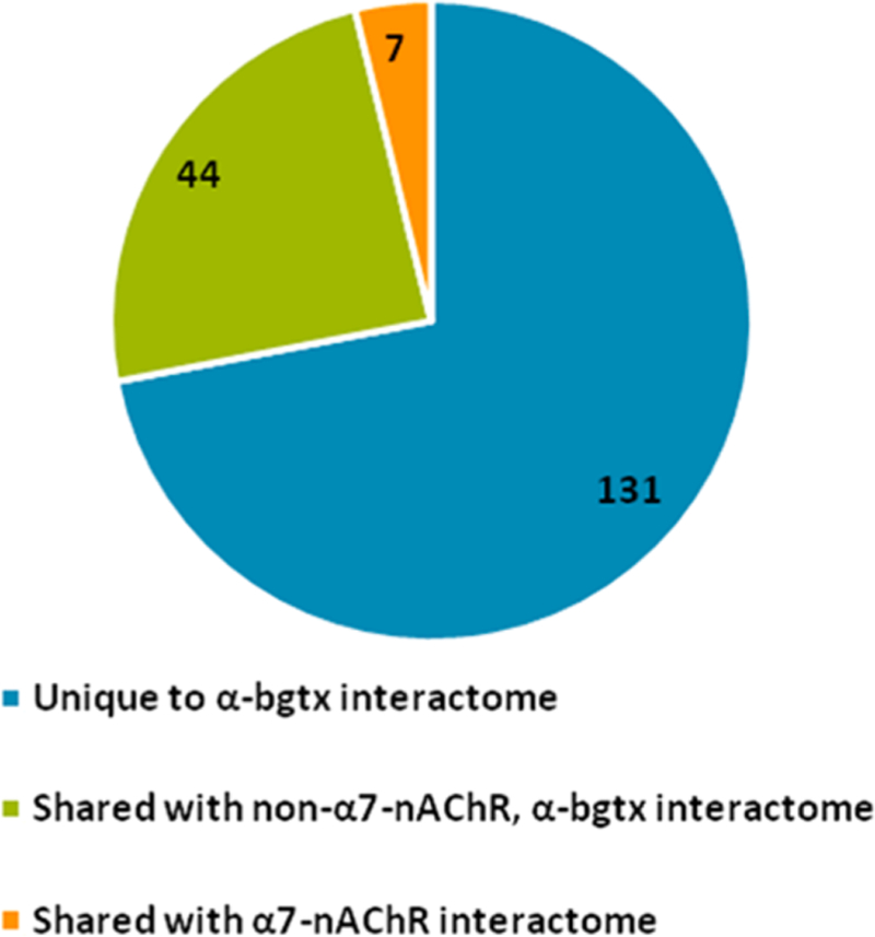
The α7-nicotinic acetylcholine receptor (α7-nAChR) is a ligand-gated ion channel that is expressed widely in vertebrates and is the principal high-affinity α-bungarotoxin (α-bgtx) binding protein in the mammalian CNS. α7-nAChRs associate with proteins that can modulate its properties. The α7-nAChR interactome is the summation of proteins interacting or associating with α7-nAChRs in a protein complex. To identify an α7-nAChR interactome in neural tissue, we isolated α-bgtx-affinity protein complexes from wild-type and α7-nAChR knockout (α7 KO) mouse whole brain tissue homogenates using α-bgtx-affinity beads. Affinity precipitated proteins were trypsinized and analyzed with an Orbitrap Fusion mass spectrometer. Proteins isolated with the α7-nAChR specific ligand, α-bgtx, were determined to be α7-nAChR associated proteins. The α7-nAChR subunit and 120 additional proteins were identified. Additionally, 369 proteins were identified as binding to α-bgtx in the absence of α7-nAChR expression, thereby identifying nonspecific proteins for α7-nAChR investigations using α-bgtx enrichment. These results expand on our previous investigations of α7-nAChR interacting proteins using α-bgtx-affinity bead isolation by controlling for differences between α7-nAChR and α-bgtx-specific proteins, developing an improved protein isolation methodology, and incorporating the latest technology in mass spectrometry. The α7-nAChR interactome identified in this study includes proteins associated with the expression, localization, function, or modulation of α7-nAChRs, and it provides a foundation for future studies to elucidate how these interactions contribute to human disease.

Keywords: Orbitrap Fusion; interactome; mass spectrometry; nAChR; nicotine; proteomics; α-bungarotoxin.


Conflict of interest statement

The authors declare no competing financial interest.

Figures

Figure 1.
Figure 1.
Murine whole brain experimental design. Solubilized protein was isolated from male (A) wild-type or (B) α7 KO mice and was incubated with Sepharose α-bgtx-affnity beads or Sepharose beads without α-bgtx (i.e., control beads). The α7-nAChR interactome was identified using FVB α7 KO extracts as α7-nAChR-specific controls, which were analyzed in parallel with FVB wild-type samples. For non-α7-nAChR, α-bgtx interactome investigations, proteins were identified from α7 KO FVB mice incubated with α-bgtx-affnity beads or control beads. For α-bgtx interactome investigations, wild-type C57Bl/6 α-bgtx-affnity enrichments were compared to C57Bl/6 extracts incubated with control beads. Protein bound to α-bgtx-affnity beads was sequentially eluted with 2 M NaCl sodium chloride and 1 M carbachol, reduced, alkylated, and digested with trypsin in-solution. The resulting peptides were analyzed with an Orbitrap Fusion mass spectrometer, and spectra were identified using the Mascot algorithm. Data were analyzed further using ProteoIQ.
Figure 2.
Figure 2.
α-bgtx-affnity immobilization conditions. (A) α-Bgtx-sensitive proteins were isolated from samples using wild-type mice and α-bgtx-conjugated affnity beads (i.e., α-bgtx beads). (B) Nonspecific interactions with α-bgtx beads were identified using α-bgtx binding enrichment in samples prepared from α7 KO mice. (C) Sepharose without conjugated α-bgtx (i.e., “control beads”) were used to control for nonspecific interactions to Sepharose. (D) Sepharose without conjugated α-bgtx (i.e., “control beads”) were used to control for nonspecific interactions to Sepharose in α7 KO mice samples.
Figure 3.
Figure 3.
Specific 125I-α-bgtx binding to solubilized protein isolated from C57Bl/6 and FVB solubilized murine brain tissue. Binding of 5 nM 125I-α-bgtx to protein immobilized on α-bgtx-affnity beads (4 mg α-bgtx per 1g Sepharose) incubated with male C57Bl/6 (A) and FVB (B) solubilized murine whole brain membrane fractions. Nonspecific binding was determined by the addition of 1 μM unlabeled α-bgtx to preparations prior to the addition of 125I-α-bgtx. Appreciable 125I-α-bgtx binding was observed in both C57Bl/6 and FVB strains (5.4 ± 0.5 and 6.4 ± 1 fmol 125I-α-bgtx per mg solubilized protein respectively). No appreciable binding was detected in either strain using control beads (A,B) or α7 KO FVB mice using α-bgtx-affnity beads (B). Three mice were used per condition, and extracts from each mouse were measured in duplicate. Significance between α-bgtx affnity pulldowns and either control bead or α-bgtx pulldowns from α7 KO was determined using a Student’s t-test. “*” denotes p < 0.05.
Figure 4.
Figure 4.
Summary of identified interactomes. Three interactomes were identified and categorized as (A) α7-nAChR, (B) non-α7-nAChR, α-bgtx, or (C) α-bgtx interactomes. High-salt wash-eluted and carbachol-eluted fractions were investigated in (A) and (B). Only carbachol-eluted proteins were investigated in (C). The high-salt washes were investigated to identify interacting proteins eluted before the addition of carbachol. An α7-nAChR subunit peptide was identified in the carbachol elutions of the α7-nAChR as well as in the α-bgtx interactomes (denoted by the green arrows). Peptides corresponding to α7-nAChR subunit were not identified in the high-salt wash fraction of the α7-nAChR interactome, nor in either of the non-α7-nAChR interactomes (red arrows). The total number of proteins identified using the defined inclusion criteria is listed on the right.
Figure 5.
Figure 5.
Cellular compartments enriched in the α7-nAChR interactome data identified using STRING. Proteins are identified by their gene name. GO Term identification numbers, false discovery rates, and individual matched protein names are included in Supplemental Tables S-2. Only matched cellular compartment ontologies <5% FDR are shown. Six ontologies described further in the text are highlighted in orange: mitochondrion, plasma membrane, neuron projection, cytoplasmic vesicle membrane, Golgi-associated vesicle, and endosome.
Figure 6.
Figure 6.
Molecular functions enriched in the α7-nAChR interactome data identified using STRING. Proteins are identified by their gene name. GO term identification numbers, false discovery rates, and individual matched protein names are included in Supplemental Tables S-4. Only matched molecular function ontologies <5% FDR are shown. Two ontologies, tau (τ)-protein kinase activity and carbohydrate derivative binding, are highlighted in orange and described further in the text.
Figure 7.
Figure 7.
Analysis of protein–protein interactions using STRING. STRING analysis (https://string-db.org/) of total identified α7-nAChR interacting proteins. 110 of 121 total proteins had suffcient information for STRING analysis. All options for active interaction sources and a minimum interaction score of 0.4 were selected. Edge colors represent known interactions from curated databases (teal), known interactions determined from experimentation (purple), predicted interactions from gene neighborhood (green), predicted interactions from gene fusions (red), or predicted interactions from gene co-occurrence (blue). Additional edge colors denote associations determined from text mining (yellow), coexpression (black), or protein homology (light blue). Node colors representing molecular functions include tau-protein kinase activity (red) and carbohydrate derivative binding (dark green). Node colors denoting cellular component include: mitochondrion (light green), plasma membrane (yellow), neuron projection (teal), cytoplasmic vesicle membrane (gold), endosome (purple), and cytoskeleton (blue).
Figure 8.
Figure 8.
Comparison of the carbachol eluted proteins from the three identified interactomes. The number of α-bgtx interacting proteins identified in the α7-nAChR interactome or the non-α7-nAChR, α-bgtx interactome. No proteins were identified in both the α7-nAChR and non-α7-nAChR, α-bgtx interactomes.

Similar articles

Cited by

References

    1. Dani JA; Bertrand D Nicotinic acetylcholine receptors and nicotinic cholinergic mechanisms of the central nervous system. Annu. Rev. Pharmacol. Toxicol 2007, 47 (1), 699–729. - PubMed
    1. Barry PH; Lynch JW Ligand-gated channels. IEEE transactions on nanobioscience 2005, 4 (1), 70–80. - PubMed
    1. Millar NS; Gotti C Diversity of vertebrate nicotinic acetylcholine receptors. Neuropharmacology 2009, 56 (1), 237–46. - PubMed
    1. Mulcahy MJ; Lester HA Granulocytes as models for human protein marker identification following nicotine exposure. J. Neurochem 2017, 142 (S2), 151–161. - PMC - PubMed
    1. Albuquerque EX; Pereira EF; Alkondon M; Rogers SW Mammalian nicotinic acetylcholine receptors: from structure to function. Physiol. Rev 2009, 89 (1), 73–120. - PMC - PubMed

Publication types

MeSH terms