. 2018 Dec 1;315(6):G966-G979.
doi: 10.1152/ajpgi.00087.2018. Epub 2018 Oct 4.

Knockout of ClC-2 reveals critical functions of adherens junctions in colonic homeostasis and tumorigenicity

Affiliations

Knockout of ClC-2 reveals critical functions of adherens junctions in colonic homeostasis and tumorigenicity

Younggeon Jin et al. Am J Physiol Gastrointest Liver Physiol. .

Abstract

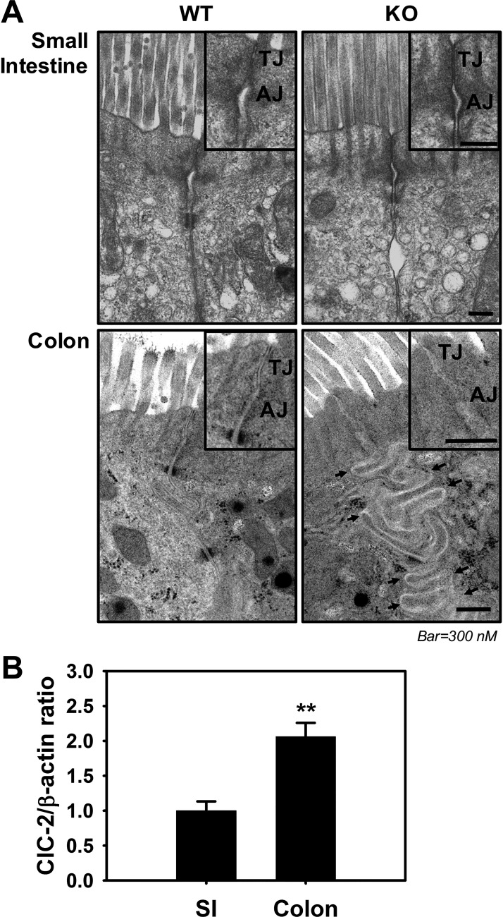
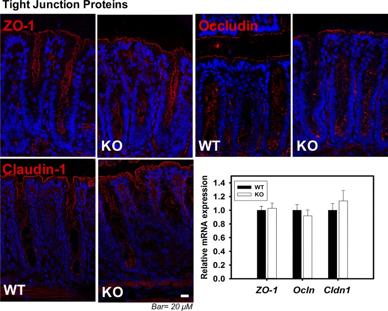
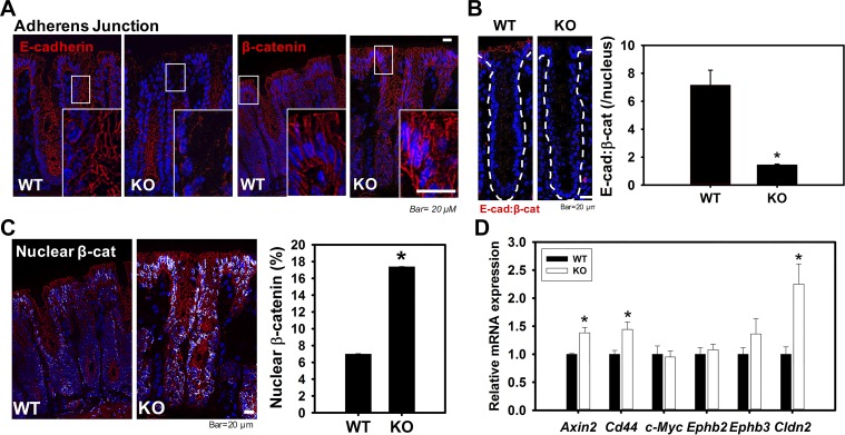
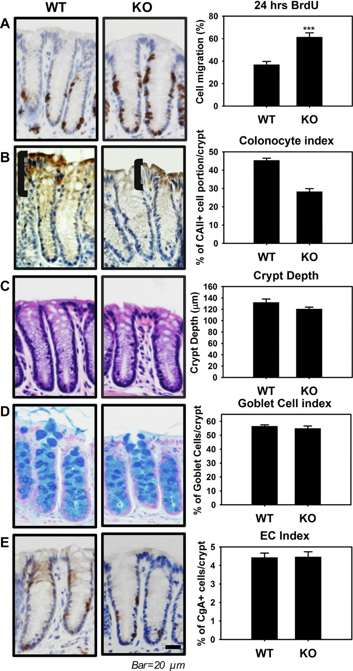
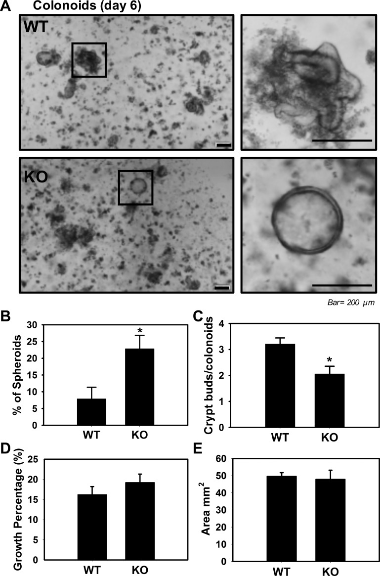
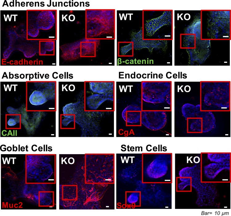
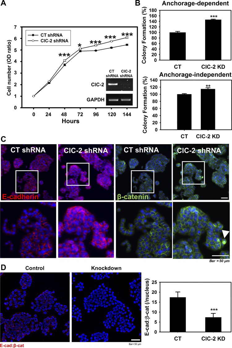
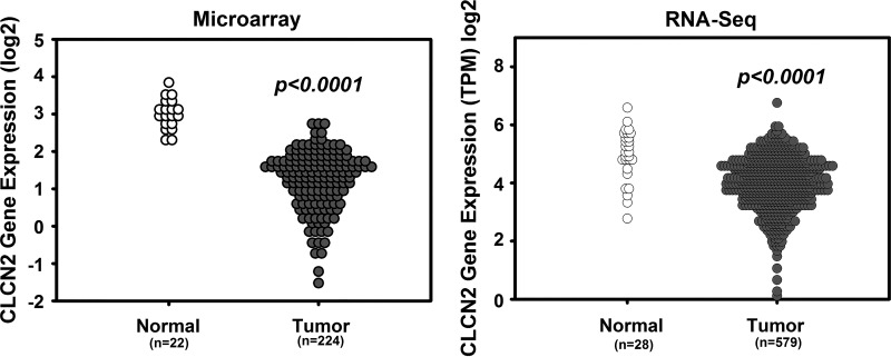
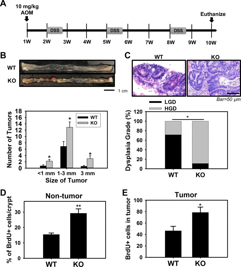
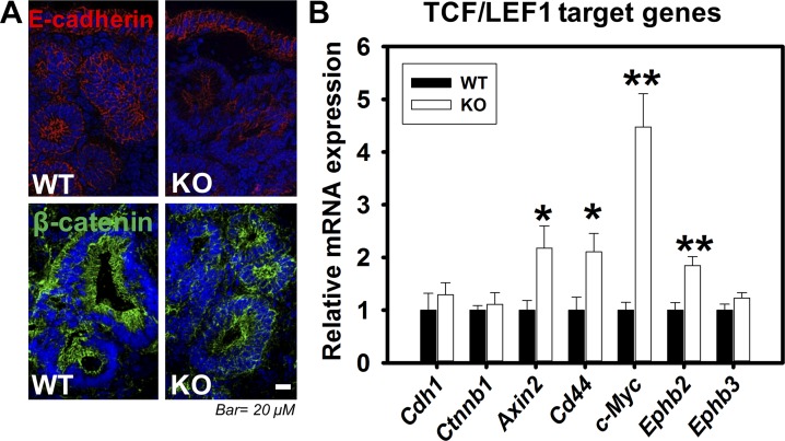
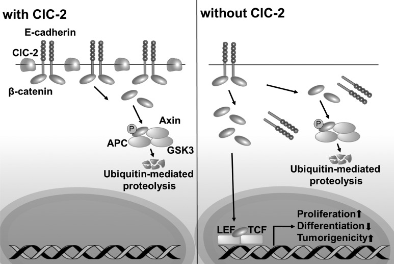
Adherens junctions (AJs), together with tight junctions (TJs), form an apical junctional complex that regulates intestinal epithelial cell-to-cell adherence and barrier homeostasis. Within the AJ, membrane-bound E-cadherin binds β-catenin, which functions as an essential intracellular signaling molecule. We have previously identified a novel protein in the region of the apical junction complex, chloride channel protein-2 (ClC-2), that we have used to study TJ regulation. In this study, we investigated the possible effects of ClC-2 on the regulation of AJs in intestinal mucosal epithelial homeostasis and tumorigenicity. Mucosal homeostasis and junctional proteins were examined in wild-type (WT) and ClC-2 knockout (KO) mice as well as associated colonoids. Tumorigenicity and AJ-associated signaling were evaluated in a murine colitis-associated tumor model and in a colorectal cancer cell line (HT-29). Colonic tissues from ClC-2 KO mice had altered ultrastructural morphology of intercellular junctions with reduced colonocyte differentiation, whereas jejunal tissues had minimal changes. Colonic crypts from ClC-2 KO mice had significantly higher numbers of less-differentiated forms of colonoids compared with WT. Furthermore, the absence of ClC-2 resulted in redistribution of AJ proteins and increased β-catenin activity. Downregulation of ClC-2 in colorectal cells resulted in significant increases in proliferation associated with disruption of AJs. Colitis-associated tumors in ClC-2 KO mice were significantly increased, associated with β-catenin transcription factor activation. The absence of ClC-2 results in less differentiated colonic crypts and increased tumorigenicity associated with colitis via dysregulation of AJ proteins and activation of β-catenin-associated signaling. NEW & NOTEWORTHY Disruption of adherens junctions in the absence of chloride channel protein-2 revealed critical functions of these junctional structures, including maintenance of colonic homeostasis and differentiation as well as driving tumorigenicity by regulating β-catenin signaling.

Keywords: adherens junction; chloride channel ClC-2; colonic homeostasis; colonic tumorigenicity; colonoids.


Figures

Fig. 1.
Fig. 1.
Role of chloride channel protein-2 (ClC-2) in ultrastructural morphology of intercellular junctions in colon. A: to evaluate the role of ClC-2 in ultrastructural morphology of intercellular junctions, images were taken using a JEOL JEM-1200EX transmission electron microscope (TEM) and Gatan ES1000 camera system of intestines from ClC-2 wild-type (WT) and knockout (KO) mice. In small intestine (top), there is no dramatic morphological change between two groups compared with colon. In WT colon, tight junctions (TJs) and adherens junctions (AJs) appeared to be more electron dense and were more closely apposed compared with ClC-2 KO colon, in which TJs and AJs were less well defined and lateral epithelial membranes were tortuously folded (black arrows) (n = 2). B: mucosa from small intestine (SI) and colon were studied for expression of ClC-2 by Western blotting. Densitometry analysis was performed to quantify expression using β-actin as a loading control. Data are presented as means ± SE (n = 4). **P < 0.01 vs. WT, Student’s t-test.
Fig. 2.
Fig. 2.
Tight junction (TJ) proteins did not show alterations between chloride channel protein-2 (ClC-2) wild-type (WT) and knockout (KO) colon. Sections of colonic tissue of ClC-2 WT and KO mice were immunolabeled for zonula occludens-1 (ZO-1), occludin, and claudin-1 (red), and the nucleus (blue). Images were taken with an Olympus IX83 Inverted Motorized Microscope and were analyzed with cellSens software. Total RNA was extracted from colonic tissues, and real-time PCR was performed with the QuantStudio 6 Flex Real-Time PCR System. ClC-2 KO mice did not show alteration of TJ protein distribution or expression (n = 6).
Fig. 3.
Fig. 3.
Chloride channel protein-2 (ClC-2) knockout (KO) mice have shown disrupted adherens junctions (AJs) with nuclear localization of β-catenin in colon compared with wild-type (WT) mice. Sections of colonic tissue of ClC-2 WT and KO mice were immunolabeled for E-cadherin and β-catenin (red), and the nucleus (blue). A: ClC-2 KO colonic crypts showed dramatic disruptions of E-cadherin in upper crypts and elevated cytosolic and nuclear distribution of β-catenin. B: proximity ligation assay (PLA) showing the interaction (red dots) between E-cadherin and β-catenin in ClC-2 KO mouse colonic crypts was significantly reduced compared with ClC-2 WT. C: nuclear β-catenin was measured using ImageJ and showed significant elevations in ClC-2 KO colon (n = 6). D: mRNA from the colonic tissues of WT and KO mice were studied for expression of T cell factor-1/lymphoid-enhancing factor-1 (TCF/LEF1) target genes by real-time PCR. The TCF/LEF1 target genes (Axin2, Cd44, and Clcn2) were significantly increased in KO colonic tissues compared with WT. Data are presented as means ± SE (n = 5–6). *P < 0.05 vs. WT, Student’s t-test.
Fig. 4.
Fig. 4.
Chloride channel protein-2 (ClC-2) knockout (KO) mice have altered colonic crypt differentiation. A: ClC-2 wild-type (WT) and KO mice were given bromodeoxyuridine (BrdU, ip) 24 h before being killed and analyzed (n = 6). The ratio of BrdU-positive cells in colonic crypts was significantly increased in ClC-2 KO mice compared with WT mice. B: immunohistochemistry (IHC) for carbonic anhydrase II (CAII) as a maker of mature colonic enterocytes. The brackets indicate the region of most highly expressed CAII in the crypts, with ClC-2 KO mice having less differentiated colonic enterocytes than WT mice. C: crypt depth in hematoxylin and eosin (H&E)-stained colonic cross sections, showing no significant difference between WT and ClC-2 KO mice. D: the no. of goblet cells per crypt was quantified using Alcian blue staining and showed no significant differences between two groups. E: the ratio of enterochromaffin cells per crypt was quantified using chromogranin A (CgA) staining and showed no significant difference between two groups. Data are presented as means ± SE (n = 6). ***P < 0.001 vs. WT, Student’s t-test.
Fig. 5.
Fig. 5.
Chloride channel protein-2 (ClC-2) loss in intestinal crypts altered formation of colonoids. Colonoid cultures from colonic crypts of ClC-2 wild-type (WT) and knockout (KO) mice harvested to form spheroids and colonoids (n = 3). Representative photos were taken (A), and quantification of spheroids (B) and crypts buds (C) was analyzed at day 6 in culture. A and B: at day 6 postculture initiation, we observed a mixed phenotype of more spheroids from ClC-2 KO colonic crypts compared with crypt-like structures in ClC-2 WT. C: the no. of buds from ClC-2 KO colonoids was significantly reduced compared with WT colonoids. D: the efficiency of colonoids from ClC-2 KO crypts (growth percentage) was not different compared with WT. E: the surface area of the colonoids was measured and showed no significant difference between two groups. Data are presented as means ± SE (n = 3). *P < 0.05 vs. WT, Student’s t-test.
Fig. 6.
Fig. 6.
Colonoids from chloride channel protein-2 (ClC-2) knockout (KO) mice had altered adherens junctions (AJ) protein distribution. Colonoids from ClC-2 wild-type (WT) and KO mice were immunolabeled for E-cadherin (red), β-catenin (green), carbonic anhydrase II (CAII, green), chromogranin A (CgA, red), Muc2 (red), and the nucleus (blue). ClC-2 KO mice had evidence of disruption of E-cadherin, elevated cytosolic and nuclear distribution of β-catenin, reduced carbonic anhydrase II (CAII), and increased Muc2.
Fig. 7.
Fig. 7.
Absence of chloride channel protein-2 (ClC-2) resulted in increased in vitro tumorigenicity and disrupted adherens junctions (AJ) proteins in HT-29 colon cancer cells. A: equal numbers of control (CT) and ClC-2 short-hairpin RNA (shRNA) cells were plated on a 96-well plate. Cell proliferation was monitored with the cell counting kit-8 (CCK-8) assay at different time points. Downregulated ClC-2 significantly increased HT-29 cell proliferation. B: in vitro tumorigenicity of the CT and ClC-2 shRNA cells was determined by a colony formation assay assessing anchorage-dependent and -independent cell populations. The tumorigenicity of ClC-2 shRNA cells was significantly increased compared with CT cells. C: ClC-2 knockdown induced disruption of AJ proteins E-cadherin and β-catenin. Arrowhead, nuclear distribution of β-catenin. D: proximity ligation assay (PLA) showing the interaction (red dots) between E-cadherin and β-catenin in ClC-2 shRNA cells was significantly reduced compared with ClC-2 wild type (WT). Data are presented as means ± SE. *P < 0.05, **P < 0.01, and ***P < 0.01 vs. CT shRNA, Student’s t-test.
Fig. 8.
Fig. 8.
Gene expression profile of chloride channel protein-2 (ClC-2, clcn2) in colorectal cancer. Gene expression data using microarray and RNA-Seq retrieved from the National Institutes of Health Cancer Genome Atlas (TCGA) project. The expression of ClC-2 was significantly reduced compared with normal colorectal tissues.
Fig. 9.
Fig. 9.
Absence of chloride channel protein-2 (ClC-2) promoted the development of colitis-associated tumors. A: protocol used for the development of colitis-associated colorectal cancer (CAC) based on the coadministration of azoxymethane (AOM, 10 mg/kg ip at day 0) and dextran sulfate sodium (DSS, 2% in 3 cycles of 5 days each at weeks 2, 5, and 8). B: absence of ClC-2 significantly elevated tumor growth and size (n = 8–9 for each group). ClC-2 knockout (KO) mice showed a significant elevation in the total number and size of colon tumors compared with wild-type (WT) mice. C: tumors were analyzed for dysplasia grade [low-grade dysplasia (LGD) and high-grade dysplasia (HGD)] and quantified based on percent of tumors demonstrating each grade. ClC-2 KO mice showed significantly increased HGD compared with WT mice, using a Fisher’s exact test. D and E: the proportion of proliferating cells in nontumor and tumor regions, quantified using bromodeoxyuridine (BrdU) staining, was significantly increased in the KO CAC mice compared with WT CAC mice. Data are presented as means ± SE. *P < 0.05 and **P < 0.01 vs. WT, Student’s t-test.
Fig. 10.
Fig. 10.
Absence of chloride channel protein-2 (ClC-2) results in activation of T cell factor-1/lymphoid-enhancing factor-1 (TCF/LEF1) target genes with disrupted adherens junctions (AJs) in colitis-associated tumors. A: sections of colon tumors from wild-type (WT) and knockout (KO) colitis-associated colorectal cancer (CAC) mice were immunolabeled for E-cadherin and β-catenin. Membranous E-cadherin (red) and β-catenin (green) were dramatically reduced in tumor regions. B: mRNA from the tumors of WT and KO mice were studied for expression of AJ proteins and TCF/LEF1 target genes by real-time PCR. mRNA expression levels of E-cadherin (Cdh1) and β-catenin (Ctnnb1) were not altered in KO CAC mice vs. WT CAC mice. However, the TCF/LEF1 target genes (Axin2, Cd44, c-Myc, and Ephb2) were significantly increased in KO CAC tumor tissues. Data are presented as means ± SE. *P < 0.05 and **P < 0.01 vs. WT, Student’s t-test.
Fig. 11.
Fig. 11.
Chloride channel protein-2 (ClC-2) has a critical function in regulation of colonic homeostasis and tumorigenicity via regulation of the stability of adherens junctions (AJs). In normal colonic mucosa, ClC-2 is associated with stability of AJ proteins to maintain colonic homeostasis. In the absence of ClC-2, colonic epithelial cells have disrupted AJ proteins E-cadherin, with internalization of β-catenin to the cytosol and subsequently the nucleus. Increased nuclear β-catenin results in alteration of colonic homeostasis and increased tumorigenicity associated with increased transcription of T cell factor-1/lymphoid-enhancing factor-1 (TCF/LEF1) target genes.

Similar articles

Cited by

References

    1. Aust DE, Terdiman JP, Willenbucher RF, Chew K, Ferrell L, Florendo C, Molinaro-Clark A, Baretton GB, Lohrs U, Waldman FM. Altered distribution of beta-catenin, and its binding proteins E-cadherin and APC, in ulcerative colitis-related colorectal cancers. Mod Pathol 14: 29–39, 2001. doi:10.1038/modpathol.3880253. - DOI - PubMed
    1. Barrett CW, Singh K, Motley AK, Lintel MK, Matafonova E, Bradley AM, Ning W, Poindexter SV, Parang B, Reddy VK, Chaturvedi R, Fingleton BM, Washington MK, Wilson KT, Davies SS, Hill KE, Burk RF, Williams CS. Dietary selenium deficiency exacerbates DSS-induced epithelial injury and AOM/DSS-induced tumorigenesis. PLoS One 8: e67845, 2013. doi:10.1371/journal.pone.0067845. - DOI - PMC - PubMed
    1. Blikslager AT, Moeser AJ, Gookin JL, Jones SL, Odle J. Restoration of barrier function in injured intestinal mucosa. Physiol Rev 87: 545–564, 2007. doi:10.1152/physrev.00012.2006. - DOI - PubMed
    1. Boivin GP, Washington K, Yang K, Ward JM, Pretlow TP, Russell R, Besselsen DG, Godfrey VL, Doetschman T, Dove WF, Pitot HC, Halberg RB, Itzkowitz SH, Groden J, Coffey RJ. Pathology of mouse models of intestinal cancer: consensus report and recommendations. Gastroenterology 124: 762–777, 2003. doi:10.1053/gast.2003.50094. - DOI - PubMed
    1. Bruun J, Kolberg M, Nesland JM, Svindland A, Nesbakken A, Lothe RA. Prognostic significance of β-catenin, E-cadherin, and SOX9 in colorectal cancer: results from a large population-representative series. Front Oncol 4: 118, 2014. doi:10.3389/fonc.2014.00118. - DOI - PMC - PubMed

Publication types