

A deep learning-based algorithm for 2-D cell segmentation in microscopy images
- PMID: 30285608
- PMCID: PMC6171227
- DOI: 10.1186/s12859-018-2375-z
A deep learning-based algorithm for 2-D cell segmentation in microscopy images
Abstract
Background: Automatic and reliable characterization of cells in cell cultures is key to several applications such as cancer research and drug discovery. Given the recent advances in light microscopy and the need for accurate and high-throughput analysis of cells, automated algorithms have been developed for segmenting and analyzing the cells in microscopy images. Nevertheless, accurate, generic and robust whole-cell segmentation is still a persisting need to precisely quantify its morphological properties, phenotypes and sub-cellular dynamics.
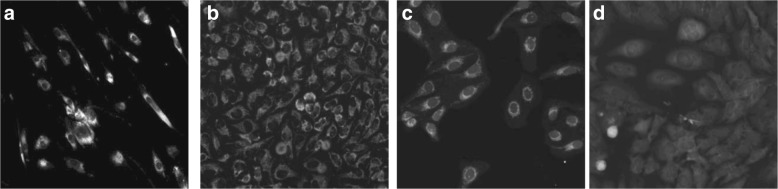
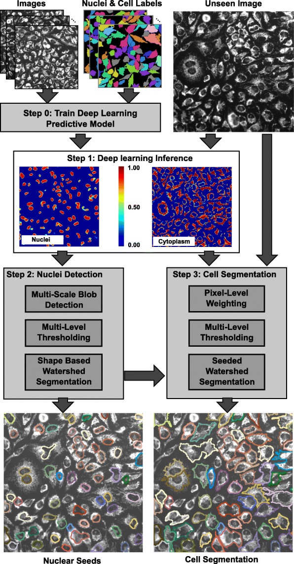
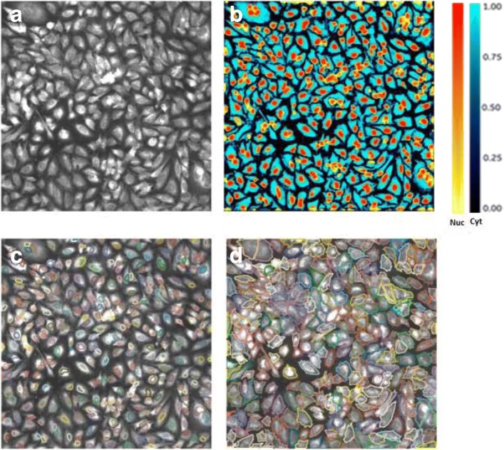
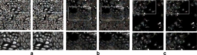
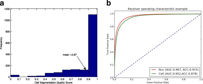
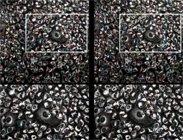
Results: We present a single-channel whole cell segmentation algorithm. We use markers that stain the whole cell, but with less staining in the nucleus, and without using a separate nuclear stain. We show the utility of our approach in microscopy images of cell cultures in a wide variety of conditions. Our algorithm uses a deep learning approach to learn and predict locations of the cells and their nuclei, and combines that with thresholding and watershed-based segmentation. We trained and validated our approach using different sets of images, containing cells stained with various markers and imaged at different magnifications. Our approach achieved a 86% similarity to ground truth segmentation when identifying and separating cells.
Conclusions: The proposed algorithm is able to automatically segment cells from single channel images using a variety of markers and magnifications.
Keywords: 2-D cells segmentation; Deep learning; Microscopy images; Watershed segmentation.
Conflict of interest statement
Ethics approval and consent to participate
Not applicable.
Consent for publication
Not applicable.
Competing interests
At the time of the submission, all of the authors of the paper were employees or contractors of General Electric. The presented algorithm was tested in a product development environment at GE Healthcare, in which several data sets were used to assess the performance of the algorithm. The datasets used for the testing and validation of the algorithm prior to the submission of the paper are provided as a supplementary material. Additional datasets were tested post submission, yet their results are beyond the scope of the current manuscript.
Publisher’s Note
Springer Nature remains neutral with regard to jurisdictional claims in published maps and institutional affiliations.
Figures
References
-
- Zhou X, Wong STC. High content cellular imaging for drug development. IEEE Signal Proc Mag. 2006;23(2):170–4. doi: 10.1109/MSP.2006.1598095. - DOI
-
- Vonesch C, Aguet F, Vonesch JL, Unser M. The colored revolution of bioimaging. IEEE Signal Proc Mag. 2006;23(3):20–31. doi: 10.1109/MSP.2006.1628875. - DOI
-
- Deshmukh BS, Mankar VH. Segmentation of microscopic images: A survey. In: 2014 International Conference on Electronic Systems, Signal Processing and Computing Technologies: 2014. p. 362–4.
-
- Meijering E. Cell Segmentation: 50 Years Down the Road. IEEE Signal Proc Mag. 2012;29(5):140–5. doi: 10.1109/MSP.2012.2204190. - DOI
MeSH terms
LinkOut - more resources
Full Text Sources
Other Literature Sources
