. 2018 Oct 3;14(1):299.
doi: 10.1186/s12917-018-1613-y.

Molecular characterization of three novel perforins in common carp (Cyprinus carpio L.) and their expression patterns during larvae ontogeny and in response to immune challenges

Affiliations

Molecular characterization of three novel perforins in common carp (Cyprinus carpio L.) and their expression patterns during larvae ontogeny and in response to immune challenges

Ting Li et al. BMC Vet Res. .

Abstract

Background: In the host immune system, perforin is a cytotoxic effector molecule that eliminate virus-infected and malignant cells. Moreover, some recent studies also imply the involvement of perforin in antibacterial immunity. Common carp (Cyprinus carpio L.), one of the most economically important fish species in China, has a high susceptibility to viruses and bacteria. Thus far, in common carp, no data are available regarding the identification and immunologic function of the perforin.

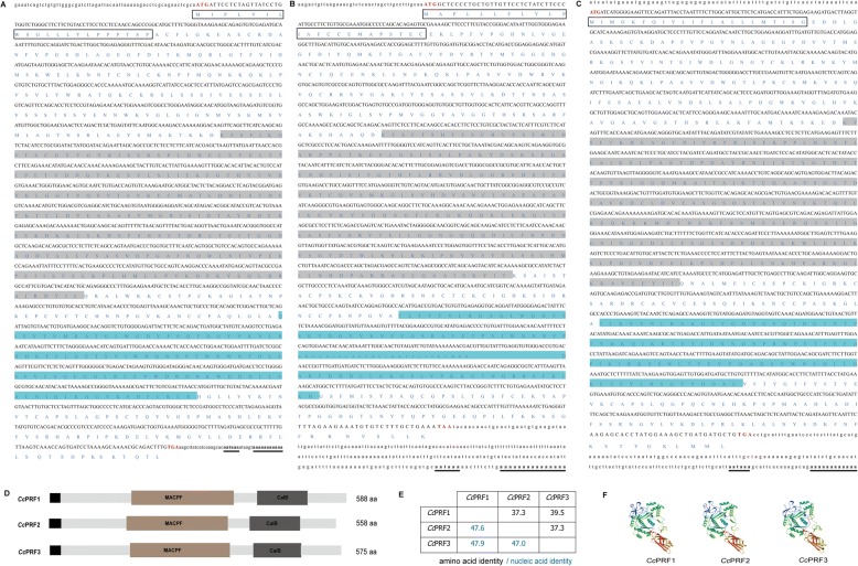
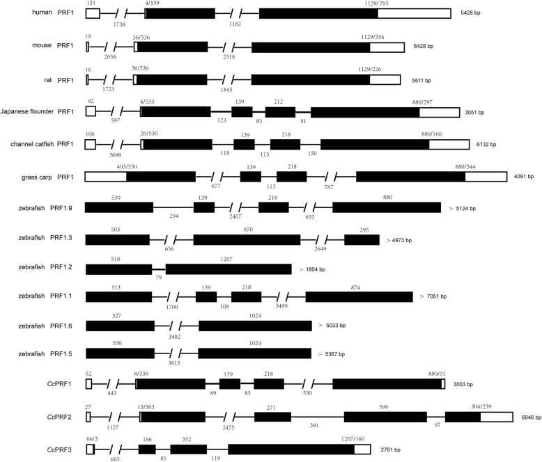
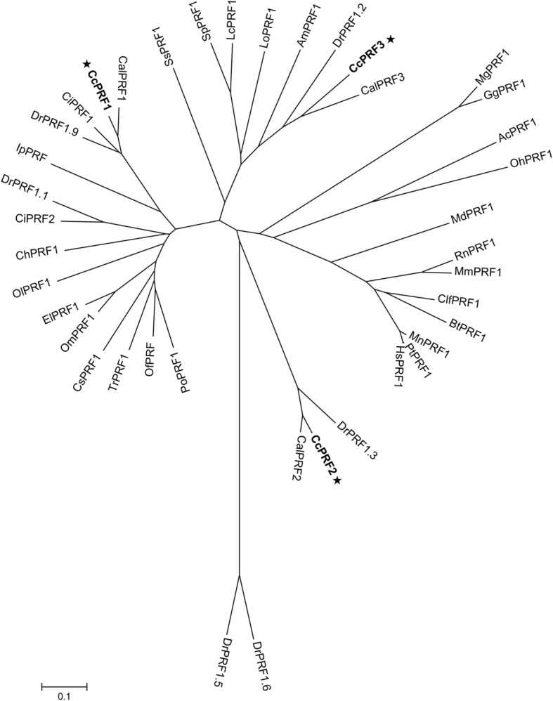
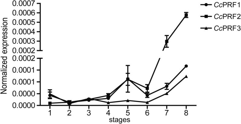
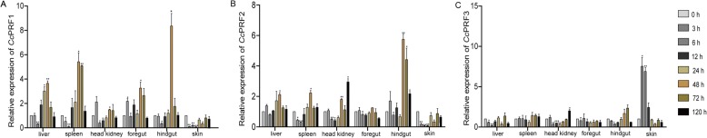
Results: In the present study, the cDNA and genomic DNA sequences of three perforin isoform genes were cloned and characterized in common carp, named CcPRF1, CcPRF2 and CcPRF3. Amino acid sequences of the three CcPRFs were quite different, with identities ranged from 37.3 to 39.5%. Phylogenetic analysis showed that three CcPRFs, each in a separate sub-branch, possessed closer evolutionary relationship with other teleost perforins, especially with cyprinid fishes, than higher vertebrates. Expression analysis revealed that each CcPRF gene was differentially expressed in all of the nine tested tissues. During larvae ontogeny, each CcPRF displayed a distinct expression pattern, while with a common expression peak at 22 days post hatching (dph). Moreover, in vivo or in vitro, after stimulation with polyI:C, LPS and Aeromonas hydrophila, each CcPRF was induced significantly, with differential expression dynamics.

Conclusions: Our findings suggest that perforin might play significant roles in larval immune system and in the immune defense of common carp against viral and bacterial pathogens. Meantime, the differential expression dynamics seem to imply possible different cellular locations or functional differences across various CcPRF isoforms.

Keywords: Common carp (Cyprinus carpio L.); Evolutionary relationship; Expression pattern; Perforin.


Conflict of interest statement

Ethics approval and consent to participate

The protocol was approved by the Animal Experimental Ethics Committee of Shandong Normal University (Permit Number: AEECSDNU2017005). Additionally, the ARRIVE (Animal Research: Reporting of in Vivo Experiments) checklist has been displayed in Additional file 10.

Consent for publication

Not applicable.

Competing interests

The authors declare that they have no competing interests.

Publisher’s Note

Springer Nature remains neutral with regard to jurisdictional claims in published maps and institutional affiliations.

Figures

Fig. 1
Fig. 1
Molecular characteristic of the three CcPRF genes. a-c cDNA and deduced amino acid sequences. CcPRF1, CcPRF2 and CcPRF3 are shown in a, b and c, respectively. The translation start codon ATG and termination codons TGA or TAA are shown in red. The signal peptides are boxed, and the MACPF and CalB domains are highlighted in light gray and blue, respectively. In addition, the polyadenylation signal (aataaa) and poly (a) tail are in bold and underlined. d Domain structures of three CcPRF proteins. The signal peptide (black), MACPF (brown) and CalB (darkgray) are depicted in different colors, and the numbers refer to the length of the amino acid sequences. e Amino acid (black) and nucleic acid (green) identities among the three CcPRFs. f 3-Dimensional protein structures were predicted using the SWISS-MODEL server, with selection of the model based on the best C-score
Fig. 2
Fig. 2
Schematic diagrams of exon-intron arrangement of various vertebrate perforin genes. Exons are shown by boxes, and introns are shown by straight or interrupted lines. The filled boxes show the coding region, while the empty ones show the untranslated region. The number of nucleotides in each exon and intron is shown above or below the corresponding element. The sequences selected are human (gene ID 5551), mouse (gene ID 18646), rat (gene ID 50669), Japanese flounder (refer to [16]), channel catfish (gene ID 108280710), grass carp (GenBank EF635861), zebrafish (gene ID: 569443, 559,849, 103,909,237, 559,384, 100,000,903, 795,573)
Fig. 3
Fig. 3
Phylogenetic analysis of perforin amino acid sequences. Phylogenetic tree is generated using the neighbor-joining (NJ) method in MEGA 6.0, and the GenBank accession numbers used are shown in Additional file 4: Table S4. Hs (Homo sapiens), Pt (Pan troglodytes), Mn (Macaca nemestrina), Rn (Rattus norvegicus), Md (Monodelphis domestica), Mm (Mus musculus), Bt (Bos Taurus), Clf (Canis lupus familiaris), Gg (Gallus gallus), Mg (Meleagris gallopavo), Oh (Ophiophagus hannah), Ac (Anolis carolinensis), El (Esox lucius), Lo (Lepisosteus oculatus), Am (Astyanax mexicanus), Sp(Stegastes partitus), Ol (Oryzias latipes), Tr (Takifugu rubripes), Om (Oncorhynchus mykiss), Lc (Larimichthys crocea), Cs (Cynoglossus semilaevis), Cal (Carassius auratus langsdorfii), Ci (Ctenopharyngodon idella), Po (Paralichthys olivaceus), Of (Oplegnathus fasciatus), Dr. (Danio rerio), Ch (Clupea harengus), Ss (Salmo salar), Ip (Ictalurus punctatus)
Fig. 4
Fig. 4
Constitutive tissue-specific expression of three CcPRF genes in healthy common carps. a Relative expression of three CcPRFs in the nine tissues (the liver, spleen, head kidney, foregut, hindgut, gill, skin, brain and muscle). b Cumulative expression and relative proportion (%) of three CcPRFs in different tissues. In each tissue, the relative proportion of CcPRF1 (bottom), CcPRF2 (middle) and CcPRF3 (top) were shown with percentage numbers. Relative gene expression level of each CcPRF was detected using real-time PCR. Amplification of S11 in each tissue was performed as an internal control (n = 4)
Fig. 5
Fig. 5
The mRNA expression patterns of three CcPRF genes during larvae ontogeny. Each CcPRF is shown with a distinct line graph. Abscissa values (1–8) are indicated as 8 developmental stages with (1) blastula stage; (2) optic primordium appearance; (3) 1 dph; (4) 2 dph; (5) 4 dph; (6) 8 dph; (7) 14 dph; (8) 22 dph. The relative expression of each CcPRF is normalized to S11 gene and displayed as the means ± SD (n = 5)
Fig. 6
Fig. 6
Gene expression of three CcPRF genes after polyI:C stimulation in vivo. CcPRF1, CcPRF2 and CcPRF3 are shown in a, b and c, respectively. The results are normalized to S11 gene. Data are presented as a fold change of the stimulated group to the un-stimulated group (denoted by 0 h) and displayed as the means ± SD (n = 3). *p < 0.05, **p < 0.01 and ***p < 0.001
Fig. 7
Fig. 7
Gene expression of three CcPRF genes after A. hydrophila stimulation in vivo. CcPRF1, CcPRF2 and CcPRF3 are shown in a, b and c, respectively. The results are normalized to S11 gene. Data are presented as a fold change of the stimulated group to the un-stimulated group (denoted by 0 h) and displayed as the means ± SD (n = 3). *p < 0.05, **p < 0.01 and ***p < 0.001
Fig. 8
Fig. 8
Gene expression of three CcPRF genes after polyI:C and LPS stimulation in vitro. a Induced expression of the CcPRFs by polyI:C in PBLs. b Induced expression of the CcPRFs by polyI:C in HKLs. c Induced expression of the CcPRFs by LPS in PBLs. d Induced expression of the CcPRFs by LPS in HKLs. Three groups of PBLs or HKLs were treated with polyI:C or LPS independently. The results are normalized to S11 gene. Data are presented as a fold change of the stimulated group to the un-stimulated group (denoted by 0 h) and displayed as the means ± SD (n = 3). *p < 0.05, **p < 0.01 and ***p < 0.001
Fig. 9
Fig. 9
Gene expression of CD8α after polyI:C and LPS stimulation in vitro. a Induced expression of CD8α by polyI:C in PBLs. b Induced expression of CD8α by polyI:C in HKLs. c Induced expression of CD8α by LPS in PBLs. d Induced expression of CD8α by LPS in HKLs. Three groups of PBLs were treated with polyI:C or LPS independently. The results are normalized to S11 gene. Data are presented as a fold change of the stimulated group to the un-stimulated group (denoted by 0 h) and displayed as the means ± SD (n = 3). *p < 0.05, **p < 0.01 and ***p < 0.001

References

    1. Kagi D, Ledermann B, Burki K, Seiler P, Odermatt B, Olsen KJ, Podack ER, Zinkernagel RM, Hengartner H. Cytotoxicity mediated by T cells and natural killer cells is greatly impaired in perforin-deficient mice. Nature. 1994;369(6475):31–37. doi: 10.1038/369031a0. - DOI - PubMed
    1. Komai-Koma M, Gilchrist DS, Xu D. Direct recognition of LPS by human but not murine CD8+ T cells via TLR4 complex. Eur J Immunol. 2009;39(6):1564–1572. doi: 10.1002/eji.200838866. - DOI - PubMed
    1. Lu CC, Wu TS, Hsu YJ, Chang CJ, Lin CS, Chia JH, Wu TL, Huang TT, Martel J, Ojcius DM, et al. NK cells kill mycobacteria directly by releasing perforin and granulysin. J Leukoc Biol. 2014;96(6):1119–1129. doi: 10.1189/jlb.4A0713-363RR. - DOI - PubMed
    1. Tokmadzic VS, Tomas MI, Sotosek S, Laskarin G, Dominovic M, Tulic V, Dordevic G, Sustic A, Mrakovcic-Sutic I. Different perforin expression in peripheral blood and prostate tissue in patients with benign prostatic hyperplasia and prostate cancer. Scand J Immunol. 2011;74(4):368–376. doi: 10.1111/j.1365-3083.2011.02569.x. - DOI - PubMed
    1. Alexander AA, Maniar A, Cummings JS, Hebbeler AM, Schulze DH, Gastman BR, Pauza CD, Strome SE, Chapoval AI. Isopentenyl pyrophosphate-activated CD56+ {gamma}{delta} T lymphocytes display potent antitumor activity toward human squamous cell carcinoma. Clin Cancer Res. 2008;14(13):4232–4240. doi: 10.1158/1078-0432.CCR-07-4912. - DOI - PMC - PubMed