. 2018 Oct 3;20(1):223.
doi: 10.1186/s13075-018-1719-4.

Blocking CD248 molecules in perivascular stromal cells of patients with systemic sclerosis strongly inhibits their differentiation toward myofibroblasts and proliferation: a new potential target for antifibrotic therapy

Affiliations

Blocking CD248 molecules in perivascular stromal cells of patients with systemic sclerosis strongly inhibits their differentiation toward myofibroblasts and proliferation: a new potential target for antifibrotic therapy

Paola Di Benedetto et al. Arthritis Res Ther. .

Abstract

Background: Fibrosis may be considered the hallmark of systemic sclerosis (SSc), the end stage triggered by different pathological events. Transforming growth factor-β (TGF-β) and platelet-derived growth factor BB (PDGF-BB) are profibrotic molecules modulating myofibroblast differentiation and proliferation, respectively. There is evidence linking CD248 with these two molecules, both highly expressed in patients with SSc, and suggesting that CD248 may be a therapeutic target for several diseases. The aim of this work was to evaluate the expression of CD248 in SSc skin and its ability to modulate SSc fibrotic process.

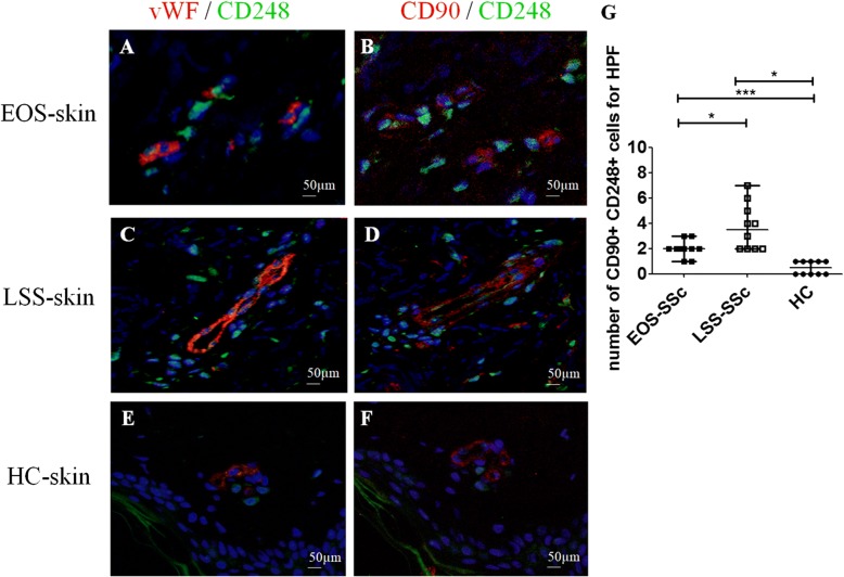
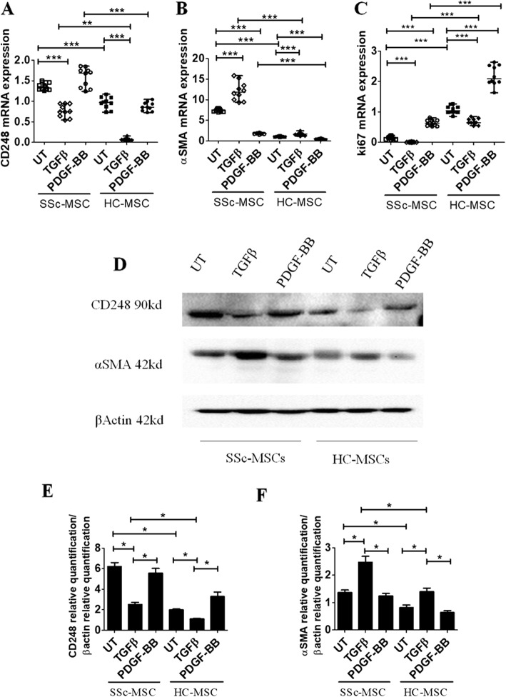
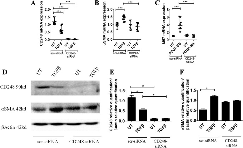
Methods: After ethical approval was obtained, skin biopsies were collected from 20 patients with SSc and 10 healthy control subjects (HC). CD248 expression was investigated in the skin, as well as in bone marrow mesenchymal stem cells (MSCs) treated with TGF-β or PDGF-BB, by immunofluorescence, qRT-PCR, and Western blotting. Finally, in SSc-MSCs, the CD248 gene was silenced by siRNA.

Results: Increased expression of CD248 was found in endothelial cells and perivascular stromal cells of SSc skin. In SSc-MSCs, the levels of CD248 and α-smooth muscle actin expression were significantly higher than in HC-MSCs. In both SSc- and HC-MSCs, PDGF-BB induced increased expression of Ki-67 when compared with untreated cells but was unable to modulate CD248 levels. After CD248 silencing, both TGF-β and PDGF-BB signaling were inhibited in SSc-MSCs.

Conclusions: CD248 overexpression may play an important role in the fibrotic process by modulating the molecular target, leading to perivascular cells differentiation toward myofibroblasts and interfering with its expression, and thus might open a new therapeutic strategy to inhibit myofibroblast generation during SSc.

Keywords: CD248; Fibrosis; Systemic sclerosis.


Conflict of interest statement

Ethics approval and consent to participate

The experiments reported in this article comply with the current ethical standard laws of Italy. All patients gave fully informed written consent approved by the institutional ethics committee.

Consent for publication

Not applicable.

Competing interests

The authors declare that they have no competing interests.

Publisher’s Note

Springer Nature remains neutral with regard to jurisdictional claims in published maps and institutional affiliations.

Figures

Fig. 1
Fig. 1
CD248 expression in skin of patients with systemic sclerosis (SSc). a, b Immunofluorescence staining of ten early-onset subset (EOS) SSc skin samples. a CD248 (green) and von Willebrand factor (vWF) (red) staining. b Consecutive section stained with CD248 (green), α-smooth muscle actin (α-SMA) (red). CD248 is expressed in endothelial cells (EC) and perivascular cells of EOS SSc skin vessels. The arrowheads show CD248+ cells localized close to microvessels. c and d Immunofluorescence staining of ten long-standing subset (LSS) SSc skin samples. c CD248 (green) and vWF (red) staining. d Consecutive section stained with CD248 (green) and α-SMA (red). CD248 is expressed in EC and perivascular cells of LSS SSc skin vessels. The arrowheads show CD248+ cells localized close to microvessels. e and f Immunofluorescence staining of ten healthy control subject (HC) skin samples. Microphotographs show (e) CD248 (green) and vWF (red) staining and (f) consecutive section stained with CD248 (green) and α-SMA (red). Weak expression of CD248 may be observed in EC and pericytes of HC skin vessels. Negative control samples were obtained by omitting the primary antibody. Original magnification × 20. g qRT-PCR of CD248 messenger RNA (mRNA) levels in ten EOS-SSc skin, ten LSS-SSc skin, and ten HC-skin samples. In SSc-skin, CD248 mRNA expression levels are always significantly higher than in HC mesenchymal stem cells. CD248 mRNA expression is significantly higher in LSS-SSc skin than in EOS-SSc skin. * p = 0.01, *** p < 0.0001
Fig. 2
Fig. 2
CD248+/CD90+ mesenchymal stem cells (MSCs) surrounding the vessels in systemic sclerosis (SSc) skin. a, b Immunofluorescence staining of ten early-onset subset (EOS) SSc skin samples. Microphotographs show (a) CD248 (green) and von Willebrand factor (vWF) (red) staining and (b) consecutive section stained with CD248 (green) and CD90 (red). c and d Immunofluorescence staining of ten long-standing subset (LSS) SSc skin samples. Microphotographs show (c) CD248 (green) and vWF (red) staining and (d) consecutive section stained with CD248 (green) and CD90 (red). e and f Immunofluorescence staining of ten healthy control subject (HC) skin samples. Microphotographs show (e) CD248 (green) and vWF (red) staining and (f) consecutive section stained with CD248 (green) and CD90 (red). Negative controls were obtained by omitting the primary antibody. Original magnification × 20. g Median number of CD90+/CD248+ cells. The number of CD90+/CD248+ cells is significantly higher in LSS-SSc skin than in EOS-SSc skin. Any dot plot is representative of the median cell count per 5 high-power fields (HPF) (× 40) for each patient. * p = 0.02, *** p = 0.0001
Fig. 3
Fig. 3
Transforming growth factor (TGF)-β and platelet-derived growth factor (PDGF)-BB effects on CD248, α-smooth muscle actin (α-SMA), and Ki-67 expression in systemic sclerosis (SSc) mesenchymal stem cells (MSCs). a qRT-PCR of CD248 messenger RNA (mRNA) levels in ten SSc-MSC (five early-onset subset [EOS] and five long-standing subset [LSS]) and ten healthy control subject (HC) MSC samples. In SSc-MSCs, CD248 mRNA expression levels are always significantly higher than in HC-MSCs. b qRT-PCR of α-SMA mRNA levels in ten SSc-MSCs (five EOS and five LSS) and ten HC-MSCs. In SSc-MSCs, the α-SMA mRNA levels are always significantly higher than in HC-MSCs. In both SSc- and HC-MSCs, TGF-β treatment induces a significant increase of α-SMA mRNA expression compared with untreated (UT) cells. On the contrary, PDGF-BB treatment induces a significant decrease of α-SMA compared with UT cells in both HC- and SSc-MSCs. c qRT-PCR of Ki-67 mRNA levels in ten SSc-MSCs (five EOS and five LSS) and ten HC-MSCs. In both SSc- and HC-MSCs, TGF-β treatment induces a significant decrease of Ki-67; on the contrary, PDGF-BB induces a significant increase of Ki-67 when compared with UT cells in both SSc- and HC-MSCs. The TGF-β isoform used is TGF-β1. Any single dot in the figure represents the median of triplicate experiments for each patient ** p = 0.0002, *** p = 0.0001. d Western blot analyses performed in four SSc-MSCs (two EOS and two LSS) and four HC SSc-MSCs confirmed the results observed by qRT-PCR analyses. Pictures are representative of all experiments. e and f Densitometric analysis of (e) CD248 protein bands and (f) α-SMA protein bands. The values were expressed as protein relative quantification/β-actin relative quantification. * p = 0.02
Fig. 4
Fig. 4
Systemic sclerosis (SSc) mesenchymal stem cells (MSCs) silenced by small interfering RNA (siRNA) CD248. a qRT-PCR of CD248 in ten SSc-MSCs (five early-onset subset [EOS] and five long-standing subset [LSS]) transfected with specific CD248-siRNA or scrambled (scr)-siRNA. Cells transfected with CD248-siRNA show a decreased expression of the CD248 gene compared with cells transfected with scr-siRNA. In SSc-MSCs treated with scr-siRNA, the transforming growth factor (TGF)-β stimulus induces a significant decrease of CD248 expression. On the contrary, in CD248-siRNA cells, TGF-β is unable to modulate CD248 mRNA expression. b qRT-PCR of α-SMA in ten SSc-MSCs (five EOS and five LSS) transfected with specific CD248-siRNA or scr-siRNA. In SSc-MSCs treated with scr-siRNA, the TGF-β stimulus induces a significant increase of α-SMA mRNA expression. On the contrary, in CD248-siRNA cells, TGF-β is unable to modulate α-SMA mRNA expression. c qRT-PCR of Ki-67 in ten SSc-MSCs (five EOS and five LSS) transfected with specific CD248-siRNA or scr-siRNA. In SSc-MSCs treated with scr-siRNA, the PDGF-BB stimulus induces a significant increase of Ki-67 mRNA expression. On the contrary, in CD248-siRNA cells, PDGF-BB is unable to modulate Ki-67 mRNA expression. The TGF-β isoform used is TGF-β1. Any single dot in the figure represents the median of triplicate experiments for each patient. *** p = 0.0001. d Western blot analyses performed in four SSc-MSCs (two EOS and two LSS) confirmed the results observed by qRT-PCR analyses. Pictures are representative of all experiments. e and f Densitometric analysis of (e) CD248 protein bands and (f) α-SMA protein bands. The values were expressed as protein relative quantification/β-actin relative quantification. * p = 0.02

Similar articles

Cited by

References

    1. Lax S, Hardie D, Wilson A, Douglas M, Anderson G, Huso D, et al. The pericyte and stromal marker CD248 (endosialin) is required for efficient lymph node expansion. Eur J Immunol. 2010;40:1884–1889. doi: 10.1002/eji.200939877. - DOI - PMC - PubMed
    1. Bagley RG, Honma N, Weber W, Boutin P, Rouleau C, Shankara S, et al. Endosialin/TEM1/CD248 is a pericyte marker of embryonic and tumor neovascularisation. Microvasc Res. 2008;76:180–188. doi: 10.1016/j.mvr.2008.07.008. - DOI - PubMed
    1. Smith SW, Eardley KS, Croft A, Nwosu J, Howie AJ, Cockwell P, et al. CD248+ stromal cells are associated with progressive chronic kidney disease. Kidney Int. 2011;80:199–207. doi: 10.1038/ki.2011.103. - DOI - PubMed
    1. Maia M, de Vriese A, Janssens T, Moons M, van Landuyt K, Tavernier J, et al. CD248 and its cytoplasmic domain: a therapeutic target for arthritis. Arthritis Rheum. 2010;62:3595–3606. doi: 10.1002/art.27701. - DOI - PubMed
    1. Rettig WJ, Garin-Chesa P, Healey JH, Su SL, Jaffe EA, Old LJ. Identification of endosialin, a cell surface glycoprotein of vascular endothelial cells in human cancer. Proc Natl Acad Sci U S A. 1992;89:10832–10836. doi: 10.1073/pnas.89.22.10832. - DOI - PMC - PubMed

MeSH terms