. 2018 Oct 1;6(1):98.
doi: 10.1186/s40425-018-0406-y.

Ibuprofen supports macrophage differentiation, T cell recruitment, and tumor suppression in a model of postpartum breast cancer

Affiliations

Ibuprofen supports macrophage differentiation, T cell recruitment, and tumor suppression in a model of postpartum breast cancer

Nathan D Pennock et al. J Immunother Cancer. .

Abstract

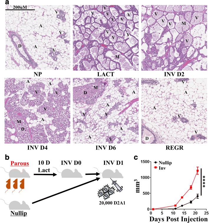
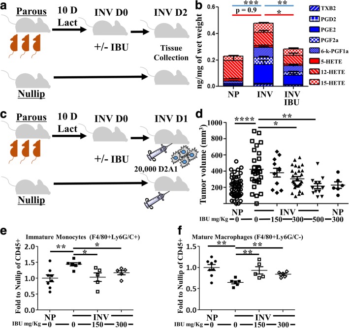
Background: Women diagnosed with breast cancer within 5 years postpartum (PPBC) have poorer prognosis than age matched nulliparous women, even after controlling for clinical variables known to impact disease outcomes. Through rodent modeling, the poor prognosis of PPBC has been attributed to physiologic mammary gland involution, which shapes a tumor promotional microenvironment through induction of wound-healing-like programs including myeloid cell recruitment. Previous studies utilizing immune compromised mice have shown that blocking prostaglandin synthesis reduces PPBC tumor progression in a tumor cell extrinsic manner. Given the reported roles of prostaglandins in myeloid and T cell biology, and the established importance of these immune cell populations in dictating tumor growth, we investigate the impact of involution on shaping the tumor immune milieu and its mitigation by ibuprofen in immune competent hosts.

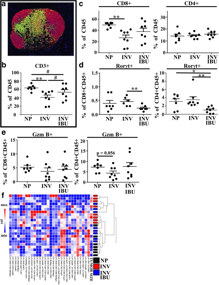
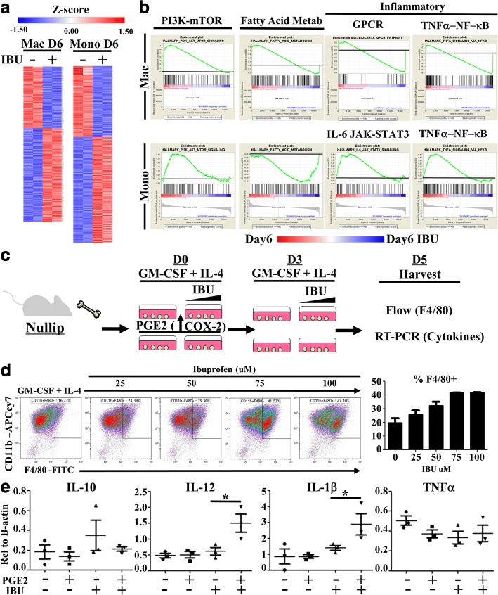
Methods: In a syngeneic (D2A1) orthotopic Balb/c mouse model of PPBC, we characterized the impact of mammary gland involution and ibuprofen treatment on the immune milieu in tumors and draining lymph nodes utilizing flow cytometry, multiplex IHC, lipid mass spectroscopy and cytokine arrays. To further investigate the impact of ibuprofen on programming myeloid cell populations, we performed RNA-Seq on in vivo derived mammary myeloid cells from ibuprofen treated and untreated involution group mice. Further, we examined direct effects of ibuprofen through in vitro bone marrow derived myeloid cell cultures.

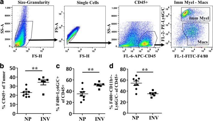
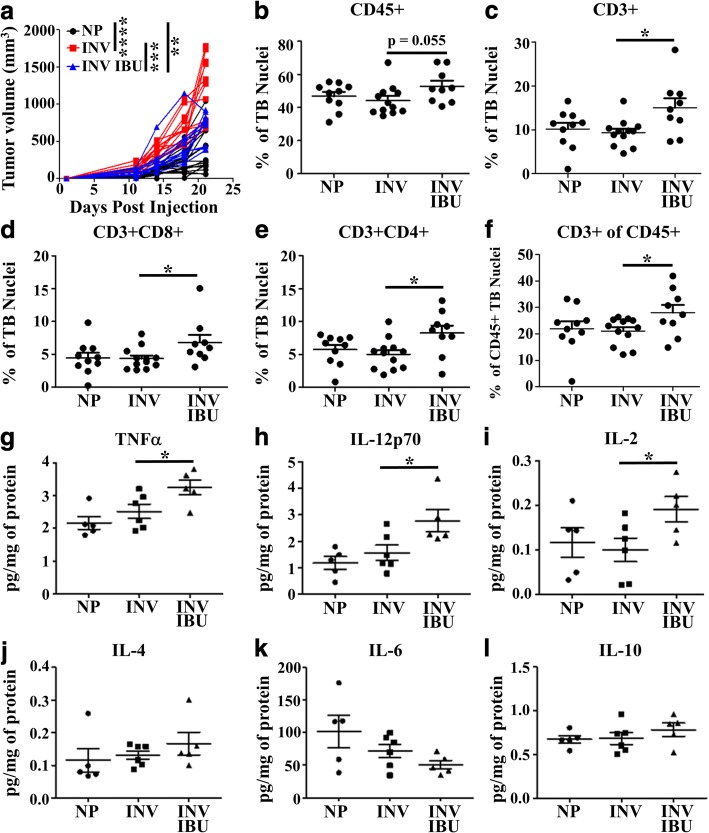
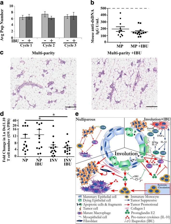
Results: Tumors implanted into the mammary involution microenvironment grow more rapidly and display a distinct immune milieu compared to tumors implanted into glands of nulliparous mice. This milieu is characterized by increased presence of immature monocytes and reduced numbers of T cells and is reversed upon ibuprofen treatment. Further, ibuprofen treatment enhances Th1 associated cytokines as well as promotes tumor border accumulation of T cells. Safety studies demonstrate ibuprofen does not impede gland involution, impact subsequent reproductive success, nor promote auto-reactivity as detected through auto-antibody and naïve T cell priming assays.

Conclusions: Ibuprofen administration during the tumor promotional microenvironment of the involuting mammary gland reduces overall tumor growth and enhances anti-tumor immune characteristics while avoiding adverse autoimmune reactions. In sum, these studies implicate beneficial prophylactic use of ibuprofen during the pro-tumorigenic window of mammary gland involution.

Keywords: Ibuprofen; Macrophages; Multiplex IHC; NSAIDs; Postpartum breast cancer; T cells; Tumor microenvironment.


Conflict of interest statement

Ethics approval and consent to participate

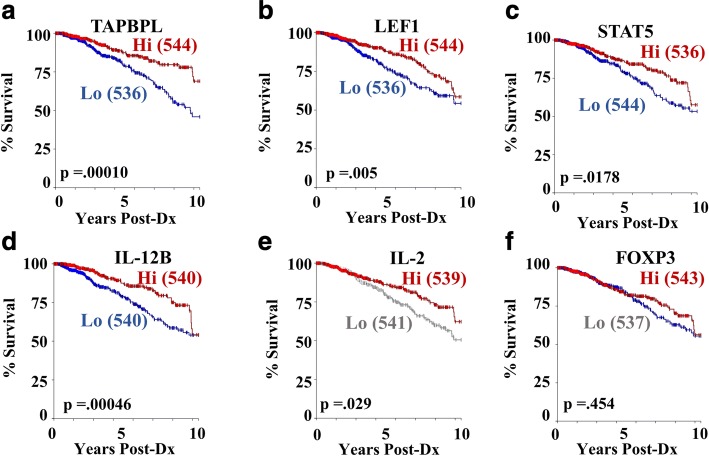
All procedures and methodologies involving mice were approved by institutional IACUC committees of the University of Colorado and Oregon Health and Science University in compliance with NIH guidelines and accepted AALAC recommendations. All represented human gene expression and outcomes data was obtained deidentified from public databases in accordance with institutional review board and health information privacy policies governing the collection and distribution of data from The Cancer Genome Atlas (TCGA).

Consent for publication

Not applicable.

Competing interests

The authors declare that they have no competing interests.

Publisher’s Note

Springer Nature remains neutral with regard to jurisdictional claims in published maps and institutional affiliations.

Figures

Fig. 1
Fig. 1
Weaning-induced mammary gland involution is a dynamic, tissue remodeling process that promotes breast cancer. a H&E sections of Balb/c mouse mammary glands across a reproductive cycle show the presence of ductal (D) and alveolar (V) epithelial structures, adipocytes (A), and milk (M) secretions within luminal spaces from nulliparous (NP), lactation (LACT), involution day two, four and six (INV D2, INV D4, and INV D6) and 6 weeks regressed (REGR) glands. b Groups of age matched Balb/c mice were bred (parous/involution, n = 8) or not (nulliparous, n = 6). Involution cohorts lactated for 10 days and pups removed to initiate synchronous mammary gland involution. One day post-weaning, 20,000 D2A1 syngeneic breast cancer line tumor cells were implanted orthotopically in the 4th mammary gland of involution or nulliparous Balb/c mice. c Tumor growth was monitored for 3 weeks before mice were euthanized for tissue collection and analyses. Determination of statistical differences between NP and INV were based upon two-tailed Student’s t-test comparison with p < 0.0001 (****)
Fig. 2
Fig. 2
Involution group tumors are enriched for immature myeloid cells. After three weeks of in vivo growth, tumors from nulliparous (NP) and involution (INV) groups were resected, digested into single cell suspensions, and myeloid populations quantified by flow cytometry. a Representative gating schema based upon scatter, single cells, CD45+, Ly6G/C and F4/80. INV compared to NP group tumors have b increased CD45+ infiltrate, c elevated immature myeloid cell/monocyte population as classified as Ly6G/C+ F4/80+ cells, and d decreased mature macrophages classified as the F4/80+ Ly6G/C– population. Determination of statistical differences between NP and INV were based upon two-tailed Student’s t-test comparison with p < 0.01 (**)
Fig. 3
Fig. 3
Seven antibody multiplex IHC analysis to characterize T cell composition of nulliparous and involution group tumors. A subset of study tumors a of representative size, growth and variation observed in the entire involution tumor cohort were evaluated for CD45, CD3, CD8, Ki67, Foxp3, Rorγt, and GzmB using multiplex IHC. b Representative image of INV group tumor where immune cell IHC signals have been pseudo-colored (all colors other than blue nuclei) illustrates an enrichment of immune cells at the tumor border (dashed white line). c Multiplex IHC signal was segmented based upon signal location relative to the tumor border (black line), with tumor border region (area between blue lines) and intratumoral regions (area inside red line) analyzed separately. d Representative image cytometry gating schema for T cell populations of interest evaluated in bulk by e hierarchical clustering of intratumoral lymphocytes and individually, showing a decrease in involution tumors of intratumoral CD3 + CD4+ T cells as proportion of f all intratumoral cells and g intratumoral CD45+ cells, with h Foxp3 + CD4+ T cells reduced as compared to all intratumoral CD3 + CD8-(putative CD4+) T cells, and i CD45 + CD3-Gzmb+ immune cells are decreased relative to intratumoral CD45+ cells during involution as well. Determination of statistical differences between nulliparous (NP) and involution (INV) were based upon two-tailed Student’s t-test comparison with p < 0.05 (*), p < 0.01 (**), and p < .001 (***)
Fig. 4
Fig. 4
Ibuprofen impairs mammary prostaglandin production, tumor promotion and immature myeloid cell frequency during involution. a INV group mice were generated by breeding, and pups nursed for 10 days (10 D). Pups were removed to initiate synchronous mammary gland involution at which time ibuprofen containing (IBU) chow (300 mg/kg chow) or normal chow was administered to INV mice. Control NP mice received normal chow only. Two days later (INV D2 for INV group mice) mammary glands were harvested and b assessed by lipid mass spectroscopy for absolute quantification of the indicated prostaglandin (blue) and leukotriene (red) family members. c The above breeding schema was repeated. Mice received the indicated dose of ibuprofen starting at initiation of involution and in age-matched NP mice. One day later, mice received an orthotopic fat pad implantation of 20,000 syngeneic D2A1 mammary epithelial tumor cells. At ~ 3 weeks post injection d tumor sizes were recorded. The tumor data are an aggregate of three independent experiments. Data were normalized between experiments by utilizing the time point where the mean tumor volume of the NP group tumors was the same for all three studies. Tumors were digested into single cell suspensions for determination of e immature monocyte and f mature macrophage content by flow cytometry. Determination of statistically significant differences were based upon two-tailed Student’s t-test between the indicated groups with p < 0.05 (*), p < 0.01 (**), p < .001 (***), p < .0001 (****)
Fig. 5
Fig. 5
Ibuprofen enhances tumor border T cell abundance and intratumoral Th1/M1 related cytokines. A subset of study tumors a representative of tumor growth from their respective groups; involution (INV), involution with 300 mg/kg ibuprofen (INV IBU) and nulliparous (NP), were stained for immune cell populations using multiplex IHC and data analyzed by image cytometry. Abundance of b CD45+, c CD3+, d CD3 + CD8+, and e CD3 + CD4 + (CD8-) as percent of total nuclei at the tumor border (TB), and composition of f CD3 + CD4 + (CD8-) relative to the CD45+, demonstrate ibuprofen effects on T cell populations at the tumor border. g-l Frozen tumor samples were subjected to cytokine level detection to determine intratumoral protein levels of g TNFa, h IL-12p70, i IL-2, j IL-4, k IL-6, and l IL-10. Determination of statistically significant differences were based upon two-tailed Student’s t-test between the indicated groups with p < 0.05 (*)
Fig. 6
Fig. 6
Ibuprofen enhances tumor draining lymph node T cell abundance and cytotoxic programming in mice. a Representative pseudo-colored composite of a T cell centric 7 antibody multiplex IHC image of tumor draining lymph node from an INV group mouse at 3wk post tumor injection (study end). CD45 is shown in red, CD3 in yellow, CD8 in green, Foxp3 in orange, GzmB in purple, Rorgt in light blue, and nuclei in blue. Quantitation of multiplex IHC signals of tumor draining lymph node to determine b CD3+ proportion of CD45+ cells, c CD3 + CD8+ and CD3 + CD4+ proportion of CD45+ cells, d Rorγt + proportion of CD8 + CD3 + CD45+ and CD4 + CD3 + CD45+ cells, and e Granzyme B (GzmB) + proportion of CD8 + CD3 + CD45+ and CD4 + CD3 + CD45+ cells. f Hierarchical clustering of lymph node multiplex IHC data show an association of evaluated parameters with tumor size and tumor cohort. Determination of statistically significant differences were based upon two-tailed Student’s t-test between the indicated groups with p < 0.05 (*) and p < 0.01 (**)
Fig. 7
Fig. 7
Involution associated macrophages and monocytes are targets of ibuprofen. Immature monocytes and macrophages from involution day 6 (D6) mice fed 300 mg/kg ibuprofen chow (+) or normal chow (−) were flow sorted based upon expression level of F4/80 and lineage negative marker. RNA was purified and subjected to whole exome RNA sequencing. a Significantly differentially expressed genes were determined based upon fold change and p-value distribution for Macrophages (Mac) and Monocytes (Mono) separately and graphically represented as relative z-score gene expression by gene (row). b Whole gene sets for macrophages (top row) or monocytes (bottom row), each with and without ibuprofen, were subjected to Gene Set Enrichment Analysis using publically available gene sets. The highly enriched gene sets for PI3K-mTOR, fatty acid metabolism, GPCR, TNFα-NFkB, and IL-6 Jak-STAT3 G protein-coupled receptor (GPCR), were selected for graphical representation. Black lines denote level of zero enrichment. Peaks or valleys that occur on the edges represent pathways differentially regulated by ibuprofen. c Bone marrow monocyte cultures were established by incubation of bone marrow cells from nulliparous animals in the presence of GM-CSF (20 ng/mL) and IL-4 (10 ng/mL) with increasing concentrations of ibuprofen for five days (D5) with or without an initial introduction of PGE2 (.92 ng/mL). Media was replenished with fresh ibuprofen where indicated on day 3 (D3). Day 5 adherent and non-adherent cells were collected for evaluation of monocyte maturation by d flow cytometry evaluation of CD11b and F4/80 expression and e RNA expression of cytokines relative to b-actin by RT-PCR. Comparisons of statistically significant differences were based upon two-tailed Student’s t-test between the indicated groups with p < 0.05 (*)
Fig. 8
Fig. 8
Ibuprofen related genes predict survival in humans TCGA Breast Cancer Data Set. Genes identified from differential expression analysis of involution macrophages, monocytes, and tumors with and without ibuprofen treatment in the above mouse studies were input as genes of interest for determination of correlation with survival in human breast cancer. The ibuprofen regulated genes a TAPBPL, b LEF1, c STAT5, d IL-12 and e IL-2 and f FOXP3 associate with better prognosis as visualized in the Kaplan-Meier plots derived from the TCGA-Breast Cancer data sets. The numbers of cases with high (Hi-red) and low (Lo-blue or gray) gene expression is indicated in parentheses
Fig. 9
Fig. 9
Ibuprofen treatment of non-tumor baring involution hosts has no observed impact on reproductive success or immune reactivity. a Numbers of pups born and successfully nursed for 10 days (start of synchronized mammary gland involution) at each cycle of breeding with (+) or without (−) 10 days of ibuprofen (IBU) after each round of weaning-induced involution (n = 10–15 mice/group). b Assessment of anti-dsDNA antibodies in the circulating maternal blood after completion of the third pregnancy/lactation/involution cycle. Maximum linear range of anti-ds-DNA detection is 1000 IgG U/mL, and gray dashed line indicates level of anti-dsDNA antibodies commonly observed in spontaneous autoimmune mouse serum. c Representative H&E sections from mice after completion of the third pregnancy/lactation/involution cycle (Multi-parity) in the presence or absence of 10 days of ibuprofen (IBU) after each round of weaning-induced involution. d Assessment of antigen specific naïve T cell activation. Assay was established as outlined in Additional file 4: Figure S4. Antigen specific T cell activation is expressed by fold change in absolute number of antigen specific Do11.10 T cells in the draining lymph node of ova injected to PBS control in nulliparous (NP) or involution (INV) mice with or without ibuprofen (IBU). Determination of statistical differences between groups was based upon one-tailed Student’s t-test according to previously published results with this assay, p < 0.05 (*). e graphical summary reported results

Similar articles

Cited by

References

    1. Azim HA, et al. Prognosis of pregnancy-associated breast cancer: a meta-analysis of 30 studies. Cancer Treat. Rev. 2012;38:834–842. - PubMed
    1. Amant F, et al. Prognosis of women with primary breast Cancer diagnosed during pregnancy: results from an international collaborative study. J Clin Oncol. 2013;31:2532–2539. doi: 10.1200/JCO.2012.45.6335. - DOI - PubMed
    1. Callihan EB, et al. Postpartum diagnosis demonstrates a high risk for metastasis and merits an expanded definition of pregnancy-associated breast cancer. Breast Cancer Res Treat. 2013;138:549–559. doi: 10.1007/s10549-013-2437-x. - DOI - PMC - PubMed
    1. McDaniel SM, et al. Remodeling of the mammary microenvironment after lactation promotes breast tumor cell metastasis. Am J Pathol. 2006;168:608–620. doi: 10.2353/ajpath.2006.050677. - DOI - PMC - PubMed
    1. Stanford JC, et al. Efferocytosis produces a prometastatic landscape during postpartum mammary gland involution. J Clin Invest. 2014;124:4737–4752. doi: 10.1172/JCI76375. - DOI - PMC - PubMed

Publication types

Substances