

Adamantinoma-like Ewing Sarcoma of the Salivary Glands: A Newly Recognized Mimicker of Basaloid Salivary Carcinomas
- PMID: 30285997
- PMCID: PMC8115302
- DOI: 10.1097/PAS.0000000000001171
Adamantinoma-like Ewing Sarcoma of the Salivary Glands: A Newly Recognized Mimicker of Basaloid Salivary Carcinomas
Abstract
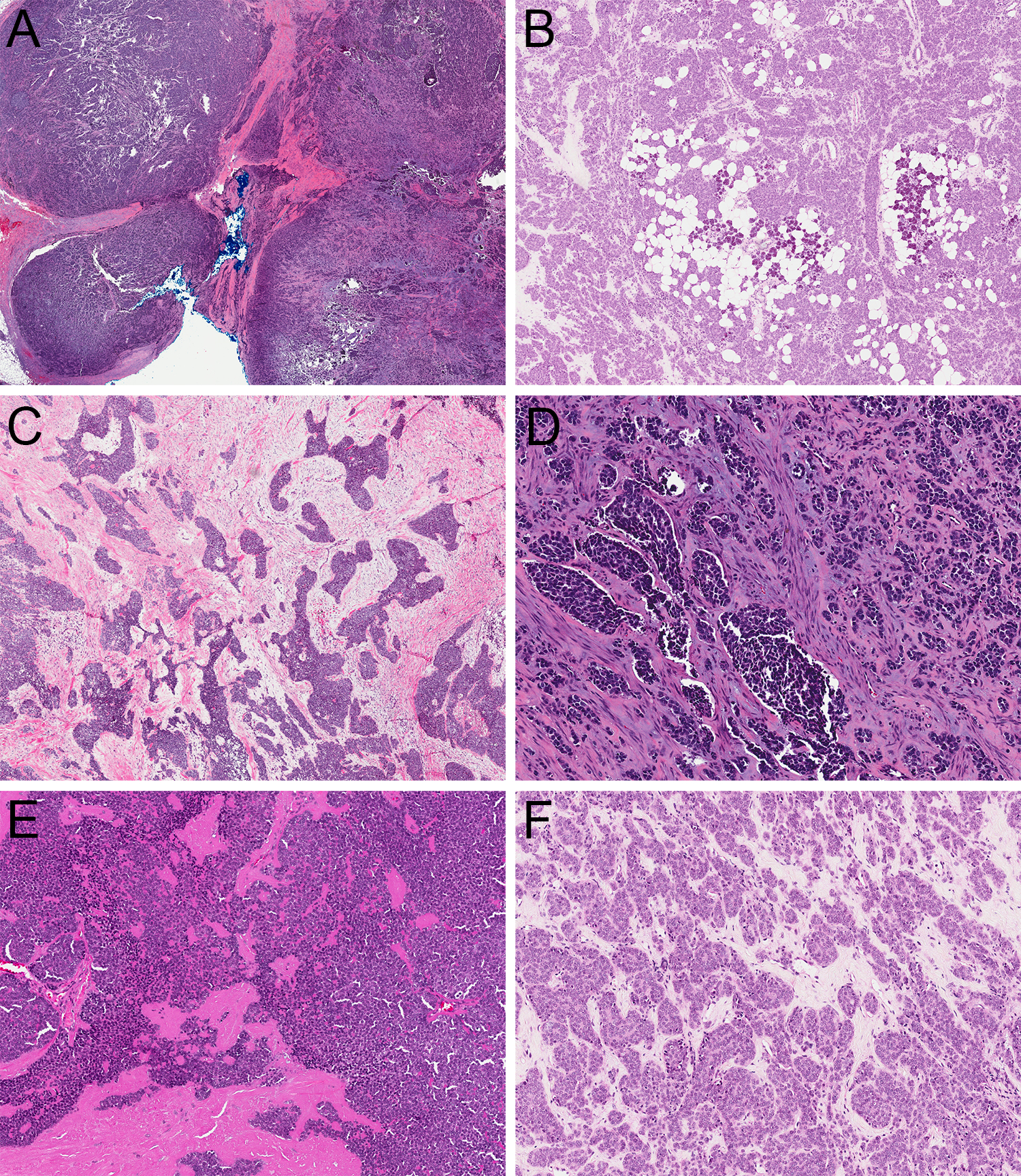
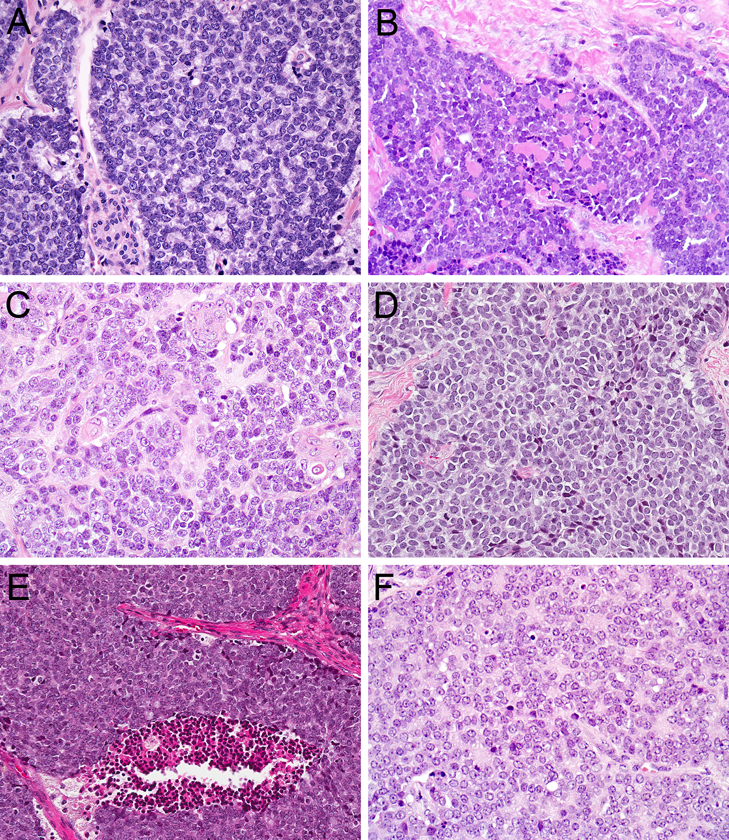
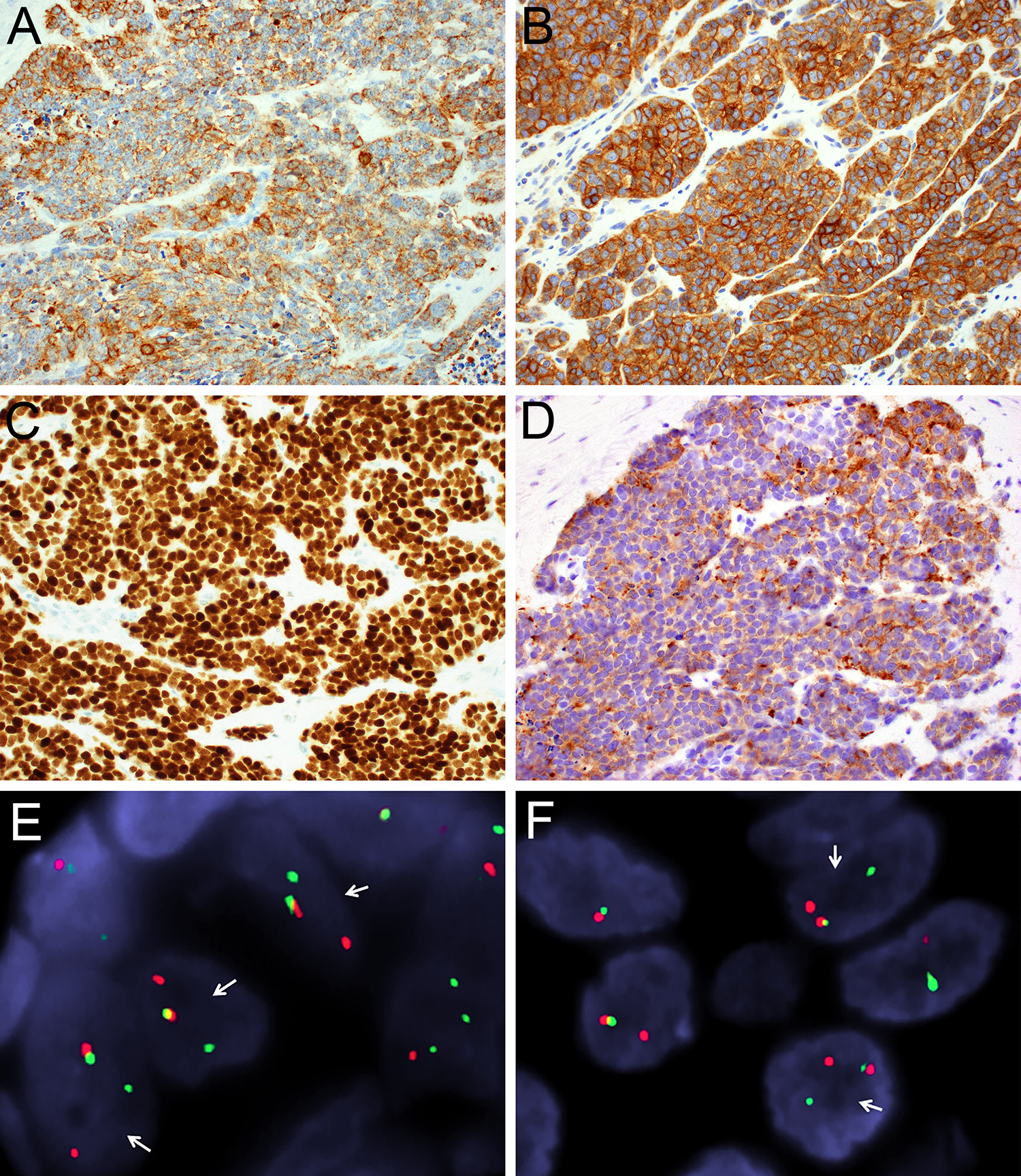
Adamantinoma-like Ewing sarcoma (ALES) is a rare tumor that demonstrates the EWSR1-FLI1 translocation characteristic of Ewing sarcoma despite overt epithelial differentiation including diffuse expression of cytokeratins and p40. Most cases of ALES described to date have occurred in the head and neck where they can mimic a wide range of small round blue cell tumors. Because distinguishing ALES from basaloid salivary gland carcinomas can be particularly difficult, we analyzed a series of 10 ALESs that occurred in the salivary glands with the aim of identifying features that allow for better recognition of this entity. The salivary ALESs included 8 parotid gland and 2 submandibular gland tumors in patients ranging from 32 to 77 years (mean: 52 y). Nine were initially misclassified as various epithelial neoplasms. Although these tumors displayed the basaloid cytology, rosette formation, infiltrative growth, and nuclear monotony characteristic of ALES, peripheral palisading and overt keratinization were relatively rare in this site. Salivary ALESs not only displayed positivity for AE1/AE3, p40, and CD99, but also demonstrated a higher proportion of synaptophysin reactivity than has been reported for nonsalivary ALESs. These morphologic and immunohistochemical findings make ALES susceptible to misclassification as various other tumors including basal cell adenocarcinoma, adenoid cystic carcinoma, squamous cell carcinoma, NUT carcinoma, large cell neuroendocrine carcinoma and myoepithelial carcinoma. Nevertheless, monotonous cytology despite highly infiltrative growth and concomitant positivity for p40 and synaptophysin can provide important clues for consideration of ALES, and identification of the defining EWSR1-FLI1 translocations can confirm the diagnosis.
Conflict of interest statement
Figures
Similar articles
-
Soft Tissue Special Issue: Adamantinoma-Like Ewing Sarcoma of the Head and Neck: A Practical Review of a Challenging Emerging Entity.Head Neck Pathol. 2020 Mar;14(1):59-69. doi: 10.1007/s12105-019-01098-y. Epub 2020 Jan 16. Head Neck Pathol. 2020. PMID: 31950471 Free PMC article. Review.
-
Adamantinoma-like Ewing Sarcoma (ALES) May Harbor FUS Rearrangements : A Potential Diagnostic Pitfall.Am J Surg Pathol. 2023 Nov 1;47(11):1243-1251. doi: 10.1097/PAS.0000000000002100. Epub 2023 Jul 27. Am J Surg Pathol. 2023. PMID: 37494548
-
Adamantinoma-like Ewing family tumors of the head and neck: a pitfall in the differential diagnosis of basaloid and myoepithelial carcinomas.Am J Surg Pathol. 2015 Sep;39(9):1267-74. doi: 10.1097/PAS.0000000000000460. Am J Surg Pathol. 2015. PMID: 26034869 Free PMC article.
-
Transcriptomic and immunophenotypic characterization of two cases of adamantinoma-like Ewing sarcoma of the thyroid gland.Histopathology. 2023 Sep;83(3):426-434. doi: 10.1111/his.14961. Epub 2023 May 17. Histopathology. 2023. PMID: 37195579
-
Adamantinoma-like Ewing sarcoma mimicking basal cell adenocarcinoma of the parotid gland: a case report and review of the literature.Head Neck Pathol. 2015 Jun;9(2):280-5. doi: 10.1007/s12105-014-0558-0. Epub 2014 Aug 1. Head Neck Pathol. 2015. PMID: 25081914 Free PMC article. Review.
Cited by
-
Top 10 Basaloid Neoplasms of the Sinonasal Tract.Head Neck Pathol. 2023 Mar;17(1):16-32. doi: 10.1007/s12105-022-01508-8. Epub 2023 Mar 16. Head Neck Pathol. 2023. PMID: 36928732 Free PMC article. Review.
-
Soft Tissue Special Issue: Adamantinoma-Like Ewing Sarcoma of the Head and Neck: A Practical Review of a Challenging Emerging Entity.Head Neck Pathol. 2020 Mar;14(1):59-69. doi: 10.1007/s12105-019-01098-y. Epub 2020 Jan 16. Head Neck Pathol. 2020. PMID: 31950471 Free PMC article. Review.
-
Fine needle biopsy of malignant tumors of the liver: a retrospective study of 624 cases from a single institution experience.Diagn Pathol. 2020 May 6;15(1):43. doi: 10.1186/s13000-020-00965-5. Diagn Pathol. 2020. PMID: 32375822 Free PMC article.
-
Adamantinoma-Like Ewing Sarcoma of the Parotid Gland: A Systematic Review.Cureus. 2025 Mar 12;17(3):e80477. doi: 10.7759/cureus.80477. eCollection 2025 Mar. Cureus. 2025. PMID: 40225424 Free PMC article. Review.
-
DNA Methylation Profiling Distinguishes Adamantinoma-Like Ewing Sarcoma From Conventional Ewing Sarcoma.Mod Pathol. 2023 Nov;36(11):100301. doi: 10.1016/j.modpat.2023.100301. Epub 2023 Aug 9. Mod Pathol. 2023. PMID: 37567448 Free PMC article.
References
-
- de Alava E, Lessnick SL, Sorenson PH. Ewing Sarcoma. In: Fletcher CD, Bridge JA, Hogendoorn PC, et al., eds. WHO Classification of Tumours of Soft Tissue and Bone. Lyon, France: International Agency for Research on Cancer; 2013:306–309.
-
- Shibuya R, Matsuyama A, Nakamoto M, et al. The combination of CD99 and NKX2.2, a transcriptional target of EWSR1-FLI1, is highly specific for the diagnosis of Ewing sarcoma. Virchows Arch. 2014;465:599–605. - PubMed
-
- Smith R, Owen LA, Trem DJ, et al. Expression profiling of EWS/FLI identifies NKX2.2 as a critical target gene in Ewing’s sarcoma. Cancer Cell. 2006;9:405–416. - PubMed
-
- Bridge JA, Fidler ME, Neff JR, et al. Adamantinoma-like Ewing’s sarcoma: genomic confirmation, phenotypic drift. Am J Surg Pathol. 1999;23:159–165. - PubMed
MeSH terms
Substances
Grants and funding
LinkOut - more resources
Full Text Sources
Medical
Miscellaneous
