. 2018 Oct 4;13(10):e0204851.
doi: 10.1371/journal.pone.0204851. eCollection 2018.

Germ cell desquamation-based testis regression in a seasonal breeder, the Egyptian long-eared hedgehog, Hemiechinus auritus

Affiliations

Germ cell desquamation-based testis regression in a seasonal breeder, the Egyptian long-eared hedgehog, Hemiechinus auritus

Diaa Massoud et al. PLoS One. .

Abstract

Testes of seasonally breeding species experience a severe functional regression before the non-breeding period, which implies a substantial mass reduction due to massive germ-cell depletion. Two alternative mechanisms of seasonal germ-cell depletion have been described in mammals, apoptosis and desquamation (sloughing), but their prevalence has not been determined yet due to reduced number of species studied. We performed a morphological, hormonal, and molecular study of the mechanism of seasonal testicular regression in males of the Egyptian long eared-hedgehog (Hemiechinus auritus). Our results show that live, non-apoptotic, germ cells are massively depleted by desquamation during the testis regression process. This is concomitant with both decreased levels of serum testosterone and irregular distribution of the cell-adhesion molecules in the seminiferous epithelium. The inactive testes maintain some meiotic activity as meiosis onset is not halted and spermatocytes die by apoptosis at the pachytene stage. Our data support the notion that apoptosis is not the major testis regression effector in mammals. Instead, desquamation appears to be a common mechanism in this class.


Conflict of interest statement

The authors have declared that no competing interests exist.

Figures

Fig 1
Fig 1
Comparisons of three morphometric parameters between summer and winter groups of H. auritus: (A) body mass, (B) testis mass and (C) seminiferous tubule diameter.
Fig 2
Fig 2
Hematoxylin‐eosin‐stained histological sections of testes (A and C) and epididymides (B and D) from H. auritus males belonging to the summer (A and B) and winter (C and D) study groups. In the summer group, the histology of the seminiferous tubules was the expected for sexually active males and the epididymides contained sperm. In contrast, testis tubules of animals collected in winter were reduced in size. They were filled with round cells resembling spermatocytes and no mature sperm was visible. In this season, the epididymal tubules are reduced in diameter and devoid of sperm. Scale bar shown in D represents 100 μm for all pictures.
Fig 3
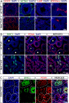
Fig 3. Immunofluorescence for several cell-type-specific molecular markers on histological sections of testes from H. auritus.
A) Analysis of the somatic cell markers SOX9 (a, e), ACTA2 (b, f), LAM (c, g) and P450scc (d, h) in active (a-d) and inactive (e-h) testes. Sertoli cells (SOX9), peritubular myoid cells (ACTA2), basal lamina (LAM) and Leydig cells (P45scc) are identified with these protein markers. B) Double immunofluorescence for the germ cell markers DMC1 and PCNA in active (a-c) and inactive (d-f) testes. In active testes, DMC1+ cells (a) were only observed in leptotene-to-early pachytene spermatocytes of the seminiferous tubules at the spermatogenic stages VII-IX (stars). PCNA expression (b) was detected in mitotic spermatogonia as well as in zygotene and pachytene spermatocytes. Both proteins co-express in zygotene and early pachytene spermatocytes of tubules at stages VII‐IX (c; stars), but not in tubules containing later pachytene spermatocytes (asterisks). In inactive testes, both markers also showed a dynamic pattern of expression similar to that of active testes, but immunoreactive cells did not present a ring-like organization and appeared clustered together (d-f). C) Higher magnification of an inactive testis showing that the luminal region of the regressed tubules was filled with either DMC1+ PCNA+ (early pachytene) or DMC1- PCNA+ (late pachytene) spermatocytes (dashed lines outline the tubular perimeter). Scale bar shown in Ah represents 50 μm for A, scale bar in Bf represents 100 μm in B and scale bar in Cd represents 50 μm in C.
Fig 4
Fig 4. Study of apoptosis (TUNEL assay) in the testes of active and inactive males of H. auritus.
A) The abundance of apoptotic cells (green) was clearly lower in active (a) than in inactive testes (b). B) Quantification of the incidence of apoptosis in active and inactive testes. C) (a-c) Double TUNEL-SOX9 immunofluorescence in inactive testes. Note that red (SOX9) and green (TUNEL) signals never colocalize, showing that Sertoli cells were not dying. (d-f) Double TUNEL-PCNA staining. All TUNEL+ cells also expressed PCNA (yellow cells in f), indicating that apoptotic cells are spermatocytes. Scale bar shown in Ab represents 100 μm for A and scale bar in Cf represents 50 μm in C.
Fig 5
Fig 5. Study of the testes of H. auritus during the regression period.
A) Hematoxylin and eosin staining of a histological section of an inactivating testis (a) and epididymis (b). During testis regression, seminiferous tubules exhibited a disorganized germinative epithelium, with primary and secondary spermatocytes occupying the lumen (see the inset in a). Round germ cells, which can be identified as primary spermatocytes (arrowhead in the inset in a), were also present in some sections of the epididymal tubule (arrowhead in the inset in b). B) TUNEL assay (green fluorescence) showing the location of several apoptotic cells. The luminal region of the seminiferous tubules in inactivating testes is mostly occupied by non-apoptotic cells. C) Expression of cell-adhesion molecules in active (a, d, g, j), inactivating (b, e, h, k) and inactive (c, f, i, l) testes. Immunofluorescence for N-CAD (a-c), β-CAT (d-f) CLDN11 (g-i) and CNX43 (j-l) showed that their expression pattern in inactive and inactivating testes is disorganized when compared with that of active testes. Scale bar shown in Ab represents 75 μm in A, scale bar in B represents 75 μm and scale bar in Cl represents 50 μm in C.
Fig 6
Fig 6. Serum testosterone concentrations in summer and winter males of H. auritus.

Similar articles

Cited by

References

    1. Neill JD. Knobil and Neill’s physiology of reproduction: Academic Press; 2006. 3296 p.
    1. Morales E, Ferrer C, Zuasti A, Garcia-Borron JC, Canteras M, Pastor LM. Apoptosis and molecular pathways in the seminiferous epithelium of aged and photoinhibited Syrian hamsters (Mesocricetus auratus). Journal of andrology. 2007;28:123–35. 10.2164/jandrol.106.000778 . - DOI - PubMed
    1. Kirkpatrick CM. The testis of the fox squirrel in relation to age and seasons. The American journal of anatomy. 1955;97:229–55. 10.1002/aja.1000970203 . - DOI - PubMed
    1. Dadhich RK, Real FM, Zurita F, Barrionuevo FJ, Burgos M, Jimenez R. Role of apoptosis and cell proliferation in the testicular dynamics of seasonal breeding mammals: a study in the Iberian mole, Talpa occidentalis. Biology of reproduction. 2010;83:83–91. 10.1095/biolreprod.109.080135 . - DOI - PubMed
    1. Pelletier RM. Cyclic formation and decay of the blood-testis barrier in the mink (Mustela vison), a seasonal breeder. The American journal of anatomy. 1986;175:91–117. 10.1002/aja.1001750109 . - DOI - PubMed

Publication types