. 2018 Oct 4;13(10):e0205189.
doi: 10.1371/journal.pone.0205189. eCollection 2018.

LC3B is not recruited along with the autophagy elongation complex (ATG5-12/16L1) at HCV replication site and is dispensable for viral replication

Affiliations

LC3B is not recruited along with the autophagy elongation complex (ATG5-12/16L1) at HCV replication site and is dispensable for viral replication

Ahmed M Fahmy et al. PLoS One. .

Abstract

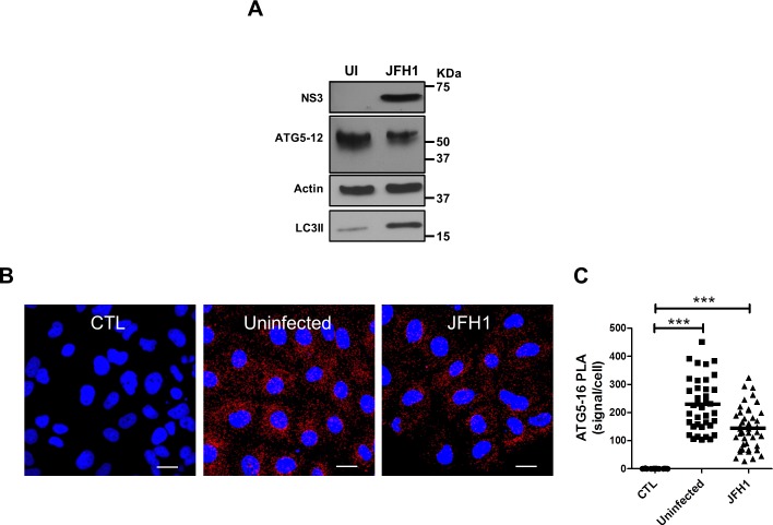
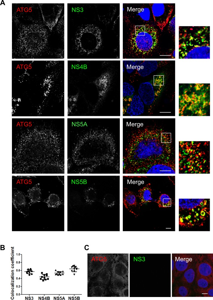
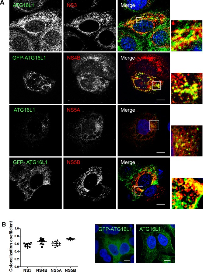
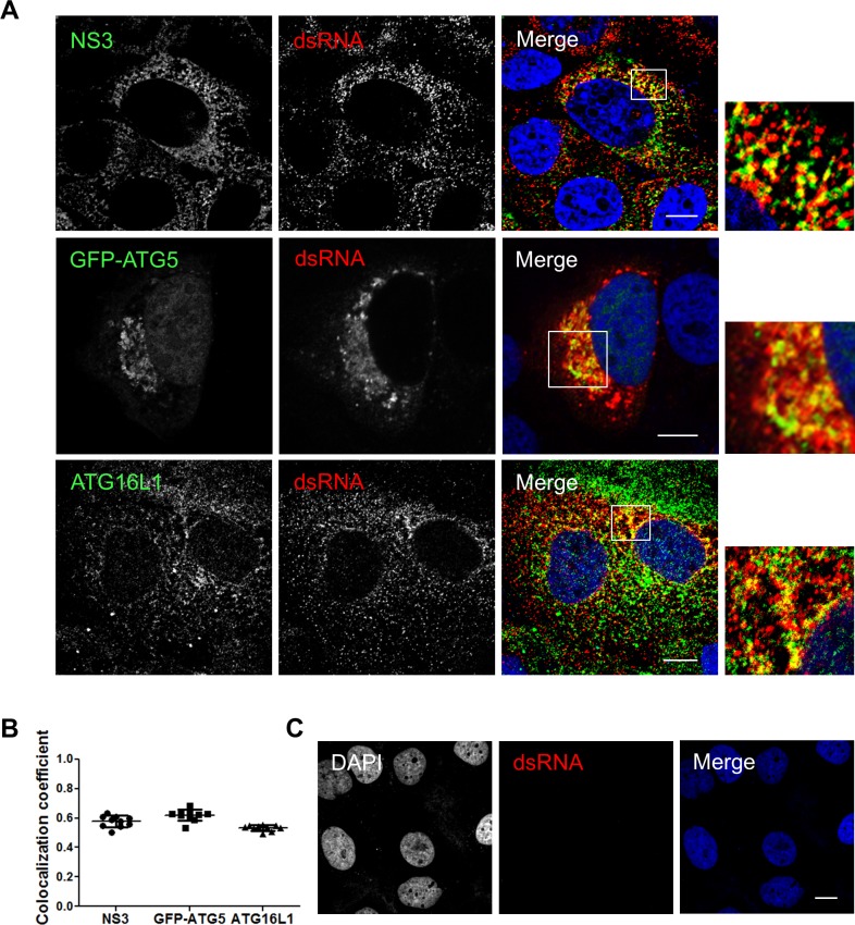
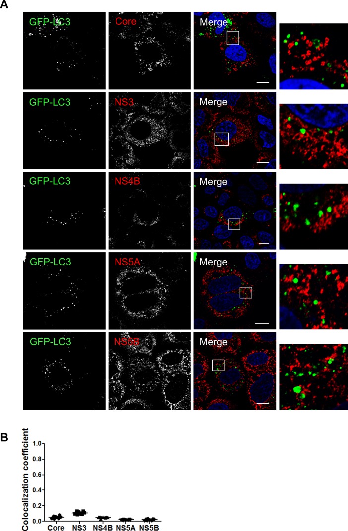
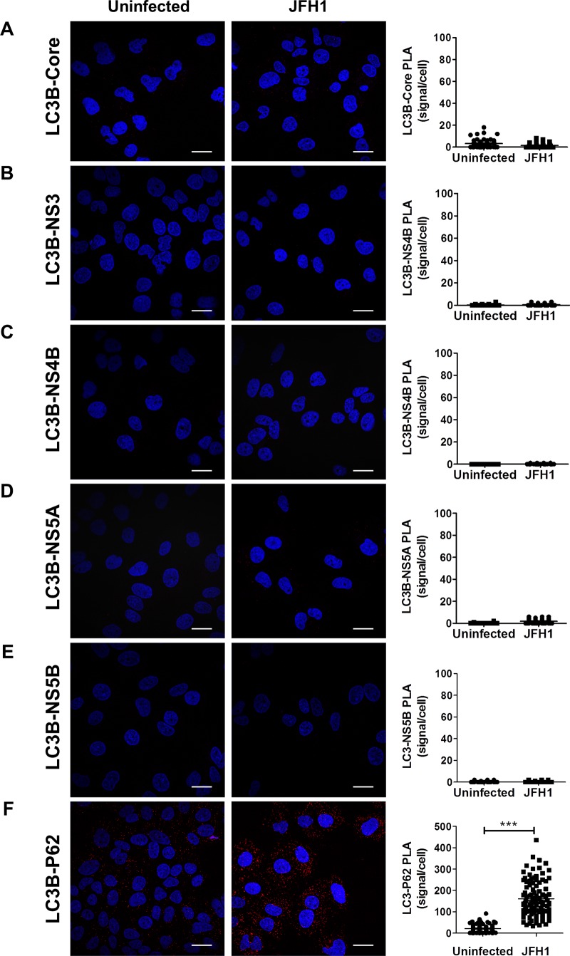
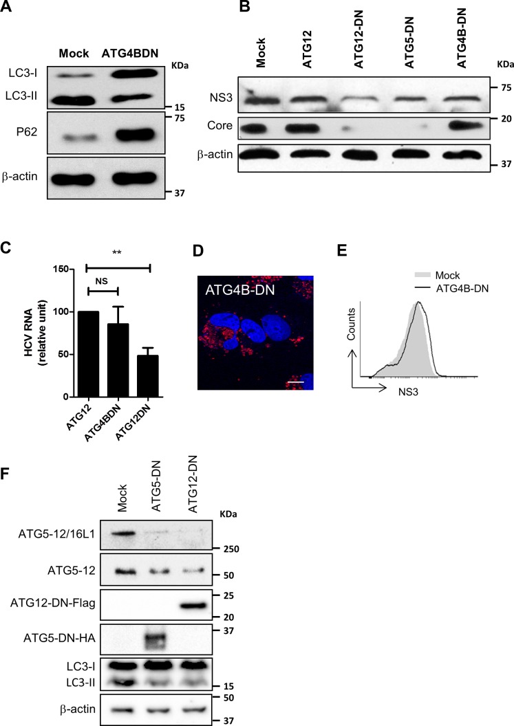
Hepatitis C virus (HCV) infection is known to induce autophagosome accumulation as observed by the typical punctate cytoplasmic distribution of LC3B-II in infected cells. Previously, we showed that viral RNA-dependent RNA polymerase (NS5B) interacts with ATG5, a major component of the autophagy elongation complex that is involved in the formation of double-membrane vesicles (DMV), and demonstrated that the autophagy elongation complex (ATG5-12/16L1) but not LC3B is required for proper membranous web formation. In this study, the colocalization and in situ interaction of all HCV replicase components with the constituent of the autophagy elongation complex and LC3B were analyzed. The results clearly show the recruitment of the elongation complex to the site of viral replication. Using in situ proximity ligation assay, we show that ATG5, but not ATG16L1, interacts with several HCV replicase components suggesting that the recruitment is directed via the ATG5-12 conjugate. Interestingly, no E3-like conjugation activity of ATG5-12/16L1 can be detected at the at HCV replication site since LC3B-II is not found along with the elongation complex at the site of viral replication. In agreement with this result, no sign of in situ interaction of LC3B with the replicase components is observed. Finally, using dominant negative forms of ATG proteins, we demonstrate that ATG5-12 conjugate, but not LC3-II formation, is critical for viral replication. Altogether, these findings suggest that although HCV needs the elongation complex for its replication, it has developed a mechanism to avoid canonical LC3-II accumulation at viral replication site.


Conflict of interest statement

The authors have declared that no competing interests exist.

Figures

Fig 1
Fig 1. Formation of the autophagy elongation complex in Huh7 cells.
A. Detection of the ATG5-12 conjugate by Western blot in mock (UI) and JFH1 infected Huh7 cells at more than 90% using an anti-ATG5 antibody. HCV infection and autophagosome accumulation were detected using anti-NS3 and anti-LC3 antibodies, respectively. β-actin represents loading control. B. In situ ATG5-12/16L1 complex formation was analyzed using PLA in JFH1-infected at more than 90% or uninfected cells. Cells were labeled for ATG5-12 and ATG16L1 using anti-ATG5 and anti-ATG16L1 respectively. CTL represents negative control lacking anti-ATG5 antibody. Nuclei were counterstained with DAPI (blue). Scale bar, 20μm. C. The frequency of PLA signals were significantly higher in both JFH1-infected and uninfected cells compared to control cells (CTL) (based on the count in 40 cells for each condition) (P<0.0001, 1way ANOVA).
Fig 2
Fig 2. Components of the HCV replicase colocalize with ATG5-12 conjugate in Huh7 cells.
A. JFH1-infected Huh7 cells at more than 90% were probed for endogenous ATG5-12 conjugate using a mouse anti-ATG5 antibody and HCV nonstructural proteins (NS3, NS4B, NS5A, and NS5B) using rabbit specific antibodies as described in the materials and methods section. The nuclei were stained with DRAQ5 (blue). Confocal microscopy images displaying subcellular localization of endogenous ATG5-12 conjugate and viral NS3, NS4A, NS5A, and NS5B in merged image panels are shown. Marked colocalization between endogenous ATG5-12 conjugate and components of the viral replicase (NS3, NS5A, and NS5B) or the membranous web (NS4B) was observed. Scale bar, 10μm. B. The average of colocalized pixels of ATG5-12 and HCV nonstructural proteins (n = 5 cells, 10 arbitrary positions) was determined. The values of overlapping fluorescence signal with HCV nonstructural proteins were calculated using Manders’ colocalization coefficient. C. Localization of ATG5-12 in uninfected cells. Uninfected Huh7 cells were immunostained for ATG5-12 using a mouse anti-ATG5 antibody. Scale bar, 10μm.
Fig 3
Fig 3. HCV nonstructural proteins colocalize with ATG12 protein in Huh7 cells.
A. Huh7 cells were infected with HCVcc and then transfected with recombinant Flag-tagged ATG12. Confocal microscopy images displaying subcellular localization of ATG12 (green) and components of the viral replicase (NS3, NS5A and NS5B) (red) in merged images are shown. Scale bar, 10μm. B. The average colocalization of ATG12 and HCV nonstructural proteins (n = 5 cells, 10 arbitrary positions) was calculated using Manders’ colocalization coefficient. C. Localization of flag-tagged ATG12 in uninfected cells. Uninfected Huh7 cells were immunostained for ATG12 using a mouse anti-flag antibody. Scale bar, 10μm.
Fig 4
Fig 4. Assessment of ATG5-12 interactions with viral proteins in infected cells as observed by proximity-ligation assay (PLA).
JFH1-infected at more than 90% or uninfected cells were fixed and processed for detection of ATG5-12-Core, ATG5-12-NS3, ATG5-12-NS4B, ATG5-12-NS5A or ATG5-12-NS5B complexes by PLA using appropriate antibodies. Nuclei were stained with DAPI (blue). Each PLA signal (red dot) indicates one interaction and were calculated as described in materials and methods section. In A, no significant difference in the frequency of PLA signals between JFH1-infected (n = 50) cells compared to uninfected controls (n = 50) indicating undetectable interaction between ATG5-12 conjugate and core. However, the incidence of PLA signals in B-E was significantly higher in JFH1-infected (n ≥23) cells compared to uninfected negative controls (N = 50) (P<0.0001, Student’s t-test) indicating complexes formation between ATG5-12 and NS3, NS4B, NS5A and NS5B. Scale bar, 20μm.
Fig 5
Fig 5. HCV nonstructural proteins colocalize with ATG16L1 in Huh7 cells.
A. Huh7 cells infected with JFH1 at more than 90% and then transfected with pGFP-ATG16L1 or immunostained for endogenous ATG16L1 using a rabbit specific antibody. Confocal microscopy images displaying subcellular localization of GFP-ATG16L1 or endogenous ATG16L1 and viral NS3, NS4B, NS5A, and NS5B are shown. Scale bar, 10μm. B. The values of overlapping fluorescence signal of ATG16L1 and GFP-ATG16L1 with HCV nonstructural proteins were calculated using Manders’ colocalization coefficient (n = 5 cells, 10 arbitrary positions). C. Uninfected Huh7 cells transfected with pGFP-ATG16L1 (left) or immunostained for endogenous ATG16L1 (right) are depicted. ATG16L1 is shown in green and the nucleus in blue (Dapi). Scale bar, 10μm.
Fig 6
Fig 6. Interaction of the autophagy elongation complex with HCV non-structural proteins is not via ATG16L1.
Using proximity-ligation assay (PLA), no in situ interaction could be observed between ATG16L1 and NS3 (A) or NS5A (B) as indicated by the frequency of PLA signals between JFH1-infected (n = 50) cells compared to uninfected controls (n = 50). Scale bar, 20μm.
Fig 7
Fig 7. The autophagy elongation complex colocalizes with HCV replicative intermediate dsRNA.
A. JFH1-infected Huh7 cells at more than 90% were immunostained for dsRNA and NS3 or ATG16L1. Alternatively, infected cells were transfected with pGFP-ATG5 and analyzed by confocal microscopy for dsRNA and GFP-ATG5. Scale bar, 10μm. B. The average of percent colocalization of NS3, GFP-ATG5 or ATG16L1 with dsRNA was determined. The values of overlapping fluorescence signals with dsRNA proteins were calculated using Manders’ colocalization coefficient (n = 5 cells, 10 arbitrary positions). C. Uninfected Huh7 cells immunostained for dsRNA. Negative staining shows specificity of dsRNA utilized in this experiment. Scale bar, 10μm.
Fig 8
Fig 8. LC3 does not colocalize with HCV proteins.
A. Huh7 cells were infected with JFH1 and then transfected with GFP-LC3. Confocal microscopy images displaying subcellular localization of GFP-LC3 and HCV core, NS3, NS4B, NS5A and NS5B are presented. Scale bar, 10μm. B. The values of overlapping fluorescence signal of GFP-LC3 with HCV nonstructural proteins were calculated using Manders’ colocalization coefficient (n = 5 cells, 10 arbitrary positions).
Fig 9
Fig 9. Assessment of LC3 interactions with viral proteins in infected cells as observed by proximity-ligation assay (PLA).
JFH1-infected at more than 90% or uninfected cells were fixed and processed for detection of LC3-Core, LC3-NS3, LC3-NS4B, LC3-NS5A, LC3-NS5B or LC3-P62 (positive control) complexes by PLA using appropriate antibodies. Nuclei were stained with DAPI (blue Interaction signals were counted as described in materials and methods. In A-E, no significant difference in the frequency of PLA signals between JFH1-infected (n = 50) cells compared to uninfected controls (n = 50) indicating undetectable interaction between LC3 and the viral proteins. F. As control, the in situ interaction between LC3 and its natural ligand P62 was measured in both infected and uninfected cells. Interestingly, the incidence of PLA signals in F was significantly higher in JFH1-infected cells compared to uninfected negative controls (N = 50) (P<0.0001, Student’s t-test). Scale bar, 20μm.
Fig 10
Fig 10. ATG5-12 conjugation but not LC3-II formation is important for the HCV lifecycle.
A. Huh7 cells were either transfected with an empty plasmid (mock) or a plasmid encoding an enzymatically inactive dominant negative form of ATG4B (ATG4B-DN). Cell lysates were analyzed by Western blot at 72 h post-transfection for LC3-I to LC3-II conversion and P62 accumulation. B. JFH1-infected Huh7 cells (>90% infected) were transfected with plasmids encoding ATG12 or the dominant negative forms of ATG5, ATG12, or ATG4B. Cell lysates of transfected cells were analyzed 72 h post-transfection for HCV core and helicase expression using anti-NS3 by western blot. β-actin was used for normalization. C. Infected Huh7 cells (>90% infected) were transfected with a plasmid encoding ATG12, ATG4B-DN or ATG12DN. Cell lysates of transfected cells were analyzed 72 h post-transfection for HCV RNA by RT-qPCR. D. Huh7 cells were transfected with pmStrawberry-ATG4BC74A (ATG4B-DN). At 48hr post transfection, immunofluorescence images were taken for ATG4BDN (red) and nucleus (blue). Scale bar, 10μm. E. Infected Huh7 cells (>90% infected) were either mock-transfected (empty plasmid) or transfected with pmStrawberry-ATG4BC74A (ATG4B-DN). Transfected cells were stained for NS3 as described in materials and methods then analyzed by flow cytometry at 72 h post-transfection for NS3 expression gating on red-fluorescent cells (pmStrawberry-ATG4B-DN positive cells). F. Huh7 cells were either transfected with an empty plasmid (mock) or a plasmid encoding a conjugation-defective dominant negative form of ATG5 (ATG5-DN) or ATG12 (ATG12-DN) tagged with HA or Flag respectively. Cell lysates were analyzed by Western blot at 72 h post-transfection for elongation complex formation (ATG5-12/16L1), ATG5-12 conjugation, and LC3-I to LC3-II conversion using anti-ATG5 and anti-LC3, respectively. The expression of ATG5-DN and ATG12-DN was verified using anti-HA and anti-Flag, respectively. β-actin was used as loading control.

Similar articles

Cited by

References

    1. Webster DP, Klenerman P, Collier J, Jeffery KJ. Development of novel treatments for hepatitis C. Lancet Infect Dis. 2009;9(2):108–17. Epub 2009/01/31. doi: S1473-3099(09)70020-9 [pii] 10.1016/S1473-3099(09)70020-9 . - DOI - PubMed
    1. Moradpour D, Penin F, Rice CM. Replication of hepatitis C virus. Nat Rev Microbiol. 2007;5(6):453–63. 10.1038/nrmicro1645 . - DOI - PubMed
    1. Wang QM, Hockman MA, Staschke K, Johnson RB, Case KA, Lu J, et al. Oligomerization and cooperative RNA synthesis activity of hepatitis C virus RNA-dependent RNA polymerase. J Virol. 2002;76(8):3865–72. 10.1128/JVI.76.8.3865-3872.2002 ; PubMed Central PMCID: PMC136118. - DOI - PMC - PubMed
    1. Sir D, Kuo CF, Tian Y, Liu HM, Huang EJ, Jung JU, et al. Replication of hepatitis C virus RNA on autophagosomal membranes. J Biol Chem. 2012;287(22):18036–43. Epub 2012/04/13. 10.1074/jbc.M111.320085 ; PubMed Central PMCID: PMC3365724. - DOI - PMC - PubMed
    1. Combs C, Brunt EM, Solomon H, Bacon BR, Brantly M, Di Bisceglie AM. Rapid development of hepatic alpha1-antitrypsin globules after liver transplantation for chronic hepatitis C. Gastroenterology. 1997;112(4):1372–5. Epub 1997/04/01. . - PubMed

Publication types

MeSH terms

Substances