. 2018 Oct 4;13(10):e0204948.
doi: 10.1371/journal.pone.0204948. eCollection 2018.

Conotoxin MVIIA improves cell viability and antioxidant system after spinal cord injury in rats

Affiliations

Conotoxin MVIIA improves cell viability and antioxidant system after spinal cord injury in rats

Karen M Oliveira et al. PLoS One. .

Abstract

This study evaluates whether intrathecal MVIIA injection after spinal cord injury (SCI) elicits neuroprotective effects. The test rats were randomly distributed into six groups- sham, placebo, MVIIA 2.5 μM, MVIIA 5 μM, MVIIA 10 μM, and MVIIA 20 μM-and were administered the treatment four hours after SCI. After the optimal MVIIA dose (MVIIA 10 μM) was defined, the best time for application, one or four hours, was analyzed. Locomotor hind limb function and side effects were assessed. Forty-eight hours after the injury and immediately after euthanasia, spinal cord segments were removed from the test rats. Cell viability, reactive oxygen species, lipid peroxidation, and glutamate release were investigated. To examine the MVIIA mechanism of action, the gene expressions of pro-apoptotic (Bax, nNOS, and caspase-3, -8, -9, -12) and anti-apoptotic (Bcl-xl) factors in the spinal cord tissue samples were determined by real-time PCR, and the activities of antioxidant enzymes were also investigated. Application of intrathecal MVIIA 10 μM four hours after SCI prompted a neuroprotective effect: neuronal death decreased (22.46%), oxidative stress diminished, pro-apoptotic factors (Bax, nNOS, and caspase-3, -8) were expressed to a lesser extent, and mitochondrial viability as well as anti-apoptotic factor (Bcl-xl) expression increased. These results suggested that MVIIA provided neuroprotection through antioxidant effects. Indeed, superoxide dismutase (188.41%), and glutathione peroxidase (199.96%), reductase (193.86%), and transferase (175.93%) expressions increased. Therefore, intrathecal MVIIA (MVIIA 10 μM, 4 h) application has neuroprotective potential, and the possible mechanisms are related to antioxidant agent modulation and to intrinsic and extrinsic apoptotic pathways.


Conflict of interest statement

The authors have declared that no competing interests exist.

Figures

Fig 1
Fig 1. Flow chart of the study design to determine the best omega-conotoxin MVIIA dose in spinal cord injury.
SHAM rats and rats subjected to SCI and injected with placebo (sterile PBS/vehicle, control, designated PLA rats) or MVIIA (designated MVIIA 2.5 μM rats, MVIIA 5 μM rats, MVIIA 10 μM rats, and MVIIA 20 μM rats).
Fig 2
Fig 2. Flow chart of the study design to determine the best application time for omega-conotoxin MVIIA in spinal cord injury.
SHAM rats and rats subjected to SCI and injected with placebo (sterile PBS/vehicle, control, designated PLA rats) or MVIIA (designated MVIIA 2.5 μM rats, MVIIA 5 μM rats, MVIIA 10 μM rats, and MVIIA 20 μM rats).
Fig 3
Fig 3. Design of the spinal segments use for each type of test.
From cranial to caudal: real-time PCR, mitochondrial viability, cell death, oxidative stress and antioxidant system.
Fig 4
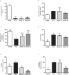
Fig 4. Different MVIIA doses effect on locomotor activity, glutamate release, mitochondrial viability, cell death, reactive oxygen species (ROS), and lipid peroxidation after spinal cord injury in Wistar rats.
Graphic representation of the results obtained for the rats subjected to dorsal laminectomy (SHAM rats, negative control) or to spinal cord injury (SCI) and subsequent injection of PBS (placebo, PLA rats) or MVIIA (MVIIA 2.5, 5, 10, and 20 μM rats) 4 h after the trauma. a) Plot of the BBB scale score (mean ± standard deviation) of deambulation in open field 24 h after surgery. The trauma groups did not differ significantly (Kruskal-Walis test, p < 0.01; SHAM, PLA, 2.5, 5, 10, and 20 μM MVIIA: 21 ± 0, 1.22 ± 0.67, 1.22 ± 0.67, 1.25 ± 0.5, 2.25 ± 1.28, and 3.8 ± 2.05, respectively). b) The glutamate concentration in the MVIIA groups was practically the same 48 h after SCI (Student-Newman-Keuls test, p > 0.05). c) Quantification of mitochondrial viability 48 h after surgery shows cell preservation in SHAM, 5 and 10 μM MVIIA in relation to PLA (100) (Student-Newman-Keuls test; PLA vs SHAM, 100% vs 223.61% ± 28.24, p < 0.01; PLA vs 5 μM MVIIA, 100% vs 183.86% ± 59.13, p < 0.05; PLA vs 10 μM MVIIA, 100% vs 180.70% ± 68.20, p < 0.05). d) The analysis of cell death 48 h after the trauma revealed significant reduction in SHAM and 10 μM MVIIA in relation to PLA (Student-Newman-Keuls test; PLA vs SHAM, 100% vs 29.46% ± 2.99, p < 0.01; PLA vs 10 μM MVIIA, 100% vs 77.43% ± 3.62, p < 0.05). e) ROS formation in SHAM, 5, 10, and 20 μM MVIIA differed from PLA rats (Student-Newman-Keuls test; PLA vs SHAM, 100% vs 59.05% ± 27.9, p < 0.05; PLA vs 5 μM MVIIA, 100% vs 16.43% ± 5.75, p < 0.01; PLA vs 10 μM MVIIA, 100% vs 22.34% ± 9.8, p < 0.01; PLA vs 20 μM MVIIA, 100% vs 16.43% ± 5.75, p < 0.01). f) The analysis of lipid peroxidation 48 h after the trauma revealed significant reduction in SHAM and 10 μM MVIIA in relation to PLA (Student-Newman-Keuls test; PLA vs SHAM, 100% vs 33.59% ± 9.14, p < 0.01; PLA vs 10 μM MVIIA, 100% vs 42.69% ± 12.38, p < 0.01). The data were normalized in relation to PLA (100). Different lowercases express statistical difference.
Fig 5
Fig 5. MVIIA effect on locomotor activity, glutamate release, mitochondrial viability, cell death, reactive oxygen species (ROS) and lipid peroxidation after spinal cord injury in Wistar rats treated with MVIIA at different times.
Graphic representation of the results obtained for the rats subjected to dorsal laminectomy (SHAM rats, negative control) or to SCI and subsequent injection of PBS (placebo, PLA rats) or MVIIA 10 μM 1 h or 4 h after surgery (MVIIA 10 μM 1h rats or MVIIA 10 μM 4h rats). a) Plot of the BBB scale score (mean ± standard deviation) of deambulation in open field 24 h after surgery. The trauma groups did not differ significantly (Kruskal-Walis test, p < 0.001; SHAM, PLA, 10 μM 1h or 10 μM 4h MVIIA: 21 ± 0, 1.33 ± 0.82, 1.67 ± 1.21, 2 ± 2.5, respectively). b) The glutamate concentration in the MVIIA groups was practically the same 48 h after SCI (Student-Newman-Keuls test, p > 0.05). c) Quantification of mitochondrial viability 48 h after surgery shows cell preservation in SHAM, 10 μM 4h MVIIA in relation to PLA (100) (Student-Newman-Keuls test; PLA vs SHAM, 100% vs 223.61% ± 28.24, p < 0.01; PLA vs 10 μM 4h MVIIA, 100% vs 180.70% ± 68.20, p < 0.05). d) The analysis of cell death 48 h after the trauma revealed significant reduction in SHAM and 10 μM 4h MVIIA in relation to PLA (Student-Newman-Keuls test; PLA vs SHAM, 100% vs 37.01% ± 15.31, p < 0.01; PLA vs 10 μM 4h MVIIA, 100% vs 77.43% ± 3.62, p < 0.05). e) ROS formation in SHAM, 10 μM 1h or 10 μM 4h MVIIA differed from PLA rats (Student-Newman-Keuls test; PLA vs SHAM, 100% vs 64.34% ± 26.9, p < 0.001; PLA vs 10 μM 1h MVIIA, 100% vs 50.32% ± 7.36, p < 0.001; PLA vs 10 μM 4h MVIIA, 100% vs 22.34% ± 9.8, p < 0.001). f) The analysis of lipid peroxidation 48 h after the trauma revealed significant reduction in SHAM and 10 μM MVIIA in relation to PLA (Student-Newman-Keuls test; PLA vs SHAM, 100% vs 33.59% ± 9.14, p < 0.01; PLA vs 10 μM MVIIA, 100% vs 42.69% ± 12.38, p < 0.01). The data were normalized in relation to PLA (100). Different lowercases express statistical difference.
Fig 6
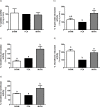
Fig 6. MVIIA effect on superoxide dismutase, catalase, glutathione peroxidase, glutathione reductase, and glutathione transferase activities 48 h after spinal cord injury.
Graphic representation of the results obtained for the rats subjected to dorsal laminectomy (SHAM rats, negative control) or to spinal cord injury and subsequent injection of PBS (placebo, PLA rats) or MVIIA 10 μM (MVIIA 10 μM rats). a) There were no catalase activity statistical differences among the groups PLA, MVIIA, and SHAM rats 48 h after SCI (Student-Newman-Keuls test, p > 0.05). b) Quantification of superoxide dismustase activity 48 h after surgery shows higher levels in MVIIA rats in relation to PLA (100) (Student-Newman-Keuls test; PLA vs MVIIA 100% vs 188.41% ± 72.05, p < 0.05). c) The analysis of glutathione peroxidase activity 48 h after the trauma revealed significant increase in MVIIA group when compared to PLA (Student-Newman-Keuls test; PLA vs MVIIA, 100% vs 199.96% ± 68.65, p < 0.01). d) Glutathione reductase activity was significantly lower in PLA rats as compared to SHAM rats (PLA vs SHAM, 100% and 215.01% ± 58.54, p < 0.01) and to MVIIA (PLA vs MVIIA, 100% vs 193.86% ± 59.39, p< 0.01, Student-Newman-Keuls test). e) Glutathione transferase activity was significantly higher in SHAM rats and MVIIA in relation to PLA (PLA vs SHAM, 100% vs 119.12% ± 8.46, p< 0.05; PLA vs MVIIA, 100% vs 175.93% vs 68.92%, p < 0.05, Student-Newman-Keuls test). The data are normalized in relation to PLA (100). Different lowercases express statistical difference.
Fig 7
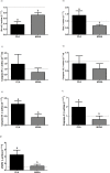
Fig 7. MVIIA effect on the relative gene expressions of Bcl-xl, Bax, caspase-9, caspase-12, caspase-8, caspase-3, and nNOS 48 h after spinal cord injury.
Mean relative gene expressions of Bcl-xl (a), Bax (b), caspase-9 (c), caspase-12 (d), caspase-8 (e), caspase-3 (f), and nNOS (g) (± standard deviation) in rats subjected to spinal cord injury associated with intrathecal administration of PBS (placebo, PLA rats) and MVIIA 10μM 4 h after SCI. Lowercases express statistical difference (a: PLA vs MVIIA, 0.72 ± 0.21 vs 0.37 ± 0.25, non-paired t test, p < 0.05; b: PLA vs MVIIA, 0.64 ± 0.18 vs 1.41 vs 0.64, Mann-Whitney test, p < 0.05; c: PLA vs MVIIA, 1.47 ± 2.21 vs 0.71 ± 0.76, Mann-Whitney test, p > 0.05; d: PLA vs MVIIA, 0.89 ± 0.94 vs 0.61 ± 0.62, non-paired t test, p > 0.05; e: PLA vs MVIIA, 0.47 ± 0.13 vs 0.17 ± 0.13, Mann-Whitney test, p < 0.05; f: PLA vs MVIIA, 1.96 ± 0.55 vs 0.60 ± 0.39, non-paired t test, p < 0.05; g: PLA vs MVIIA, 4.51 ± 2.77 vs 1.22 ± 1.14 non-paired t test, p < 0.05).

Similar articles

Cited by

References

    1. Oyinbo CA. Secondary injury mechanisms in traumatic spinal cord injury: a nugget of this multiply cascade. Acta Neurobiol Exp. 2011; 71:281–299. . - PubMed
    1. Jia X, Kowalski RG, Sciubba DM, Geocadin RG. Critical care of traumatic spinal cord injury. J Intensiv Care Med. 2003; 28:12–23. 10.1177/0885066611403270. - DOI - PubMed
    1. Eckert MJ, Martin MJ. Trauma: spinal cord injury. Surg Clin N Am. 2017; 97:1031–1045. 10.1016/j.suc.2017.06.008 - DOI - PubMed
    1. Carlson GD, Gorden C. Current developments in spinal cord injury research. Spine J. 2002; 2:116–128. 10.1016/S1529-9430(01)00029-8. - DOI - PubMed
    1. Hall ED, Springer JE. Neuroprotection and acute spinal cord injury: a reappraisal. NeuroRx: J Am Soc Exp NeuroTher. 2004; 1:80–100. 10.1602/neurorx.1.1.80. - DOI - PMC - PubMed

Publication types

MeSH terms