

A control mechanism for intra-mural peri-arterial drainage via astrocytes: How neuronal activity could improve waste clearance from the brain
- PMID: 30286191
- PMCID: PMC6171921
- DOI: 10.1371/journal.pone.0205276
A control mechanism for intra-mural peri-arterial drainage via astrocytes: How neuronal activity could improve waste clearance from the brain
Abstract
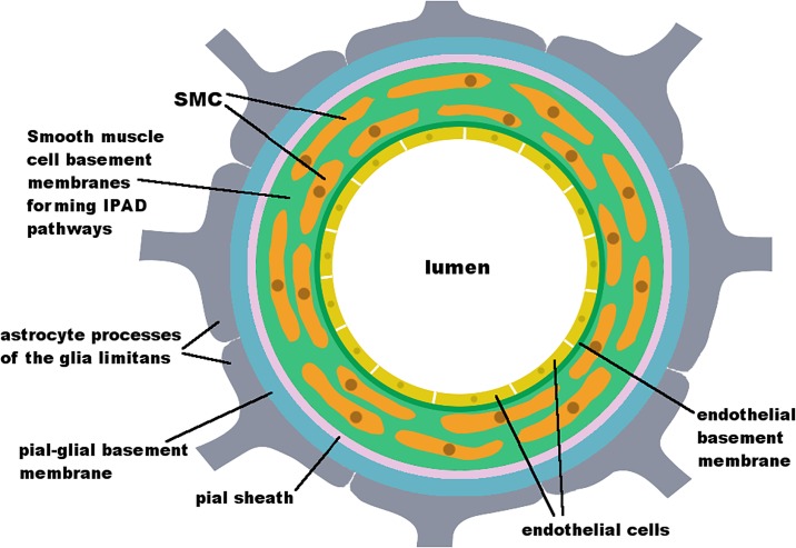
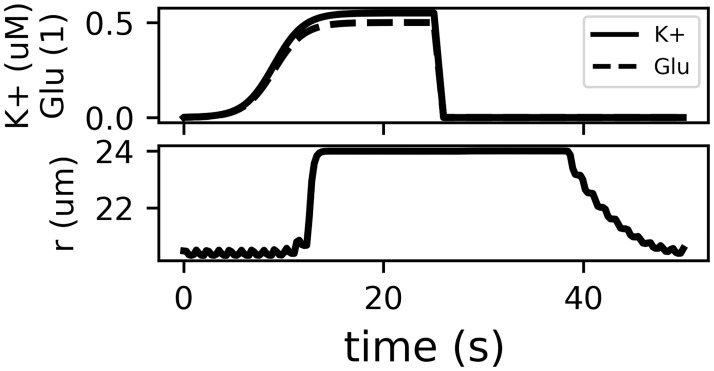
The mechanisms behind the clearance of soluble waste from deep within the parenchyma of the brain remain unclear. Experimental evidence reveals that one pathway for clearance of waste, termed intra-mural peri-arterial drainage (IPAD), is the rapid drainage of interstitial fluid along basement membranes (BM) of the smooth muscle cells of cerebral arteries; failure of IPAD is closely associated with the pathology of Alzheimer's disease (AD), but its driving mechanism remains unclear. We have previously shown that arterial pulsations generated by the heart beat are not strong enough to drive IPAD. Here we present computational evidence for a mechanism for clearance of waste from the brain that is driven by functional hyperaemia, that is, the dilatation of cerebral arterioles as a consequence of increased nutrient demand from neurons. This mechanism is based on our model for the flow of fluid through the vascular BM. It accounts for clearance rates observed in mouse experiments, and aligns with pathological observations and recommendations to lower the individual risk of AD, such as mental and physical activity. Thus, our neurovascular hypothesis should act as the new working hypothesis for the driving force behind IPAD.
Conflict of interest statement
The authors have declared that no competing interests exist.
Figures


Similar articles
-
Interstitial fluid drainage is impaired in ischemic stroke and Alzheimer's disease mouse models.Acta Neuropathol. 2013 Sep;126(3):353-64. doi: 10.1007/s00401-013-1145-2. Epub 2013 Jul 2. Acta Neuropathol. 2013. PMID: 23818064 Free PMC article.
-
Solutes, but not cells, drain from the brain parenchyma along basement membranes of capillaries and arteries: significance for cerebral amyloid angiopathy and neuroimmunology.Neuropathol Appl Neurobiol. 2008 Apr;34(2):131-44. doi: 10.1111/j.1365-2990.2007.00926.x. Epub 2008 Jan 16. Neuropathol Appl Neurobiol. 2008. PMID: 18208483
-
Cerebral amyloid angiopathy: pathogenesis and effects on the ageing and Alzheimer brain.Neurol Res. 2003 Sep;25(6):611-6. doi: 10.1179/016164103101202057. Neurol Res. 2003. PMID: 14503015 Review.
-
Cerebrovascular Smooth Muscle Cells as the Drivers of Intramural Periarterial Drainage of the Brain.Front Aging Neurosci. 2019 Jan 23;11:1. doi: 10.3389/fnagi.2019.00001. eCollection 2019. Front Aging Neurosci. 2019. PMID: 30740048 Free PMC article.
-
Lymphatic drainage of the brain and the pathophysiology of neurological disease.Acta Neuropathol. 2009 Jan;117(1):1-14. doi: 10.1007/s00401-008-0457-0. Epub 2008 Nov 11. Acta Neuropathol. 2009. PMID: 19002474 Review.
Cited by
-
Reproducibility of diffusion tensor image analysis along the perivascular space (DTI-ALPS) for evaluating interstitial fluid diffusivity and glymphatic function: CHanges in Alps index on Multiple conditiON acquIsition eXperiment (CHAMONIX) study.Jpn J Radiol. 2022 Feb;40(2):147-158. doi: 10.1007/s11604-021-01187-5. Epub 2021 Aug 14. Jpn J Radiol. 2022. PMID: 34390452 Free PMC article.
-
Neurodegeneration and inflammation crosstalk: Therapeutic targets and perspectives.IBRO Neurosci Rep. 2022 Dec 16;14:95-110. doi: 10.1016/j.ibneur.2022.12.003. eCollection 2023 Jun. IBRO Neurosci Rep. 2022. PMID: 37388502 Free PMC article.
-
The lymphatic drainage systems in the brain: a novel target for ischemic stroke?Neural Regen Res. 2023 Mar;18(3):485-491. doi: 10.4103/1673-5374.346484. Neural Regen Res. 2023. PMID: 36018151 Free PMC article. Review.
-
Relationship between Parasagittal Perivenous Cysts and Leakage of Gadolinium-based Contrast Agents into the Subarachnoid Space around the Cortical Veins after Intravenous Administration.Magn Reson Med Sci. 2021 Sep 1;20(3):245-252. doi: 10.2463/mrms.mp.2020-0062. Epub 2020 Jul 14. Magn Reson Med Sci. 2021. PMID: 32669512 Free PMC article.
-
Arterial vasodilation drives convective fluid flow in the brain: a poroelastic model.Fluids Barriers CNS. 2022 May 15;19(1):34. doi: 10.1186/s12987-022-00326-y. Fluids Barriers CNS. 2022. PMID: 35570287 Free PMC article.
References
-
- Carare RO, Bernardes-Silva M, Newman TA, Page AM, Nicoll JAR, Perry VH, et al. Solutes, but not cells, drain from the brain parenchyma along basement membranes of capillaries and arteries: Significance for cerebral amyloid angiopathy and neuroimmunology. Neuropathology and Applied Neurobiology. 2008;34(2):131–144. 10.1111/j.1365-2990.2007.00926.x - DOI - PubMed
Publication types
MeSH terms
Substances
LinkOut - more resources
Full Text Sources
Medical
