. 2018 Oct 16;115(8):1445-1456.
doi: 10.1016/j.bpj.2018.09.002. Epub 2018 Sep 13.

Prediction of the Closed Conformation and Insights into the Mechanism of the Membrane Enzyme LpxR

Affiliations

Prediction of the Closed Conformation and Insights into the Mechanism of the Membrane Enzyme LpxR

Graham M Smeddle et al. Biophys J. .

Abstract

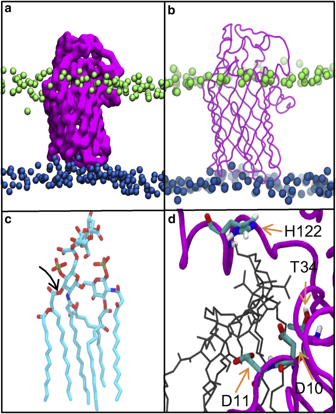
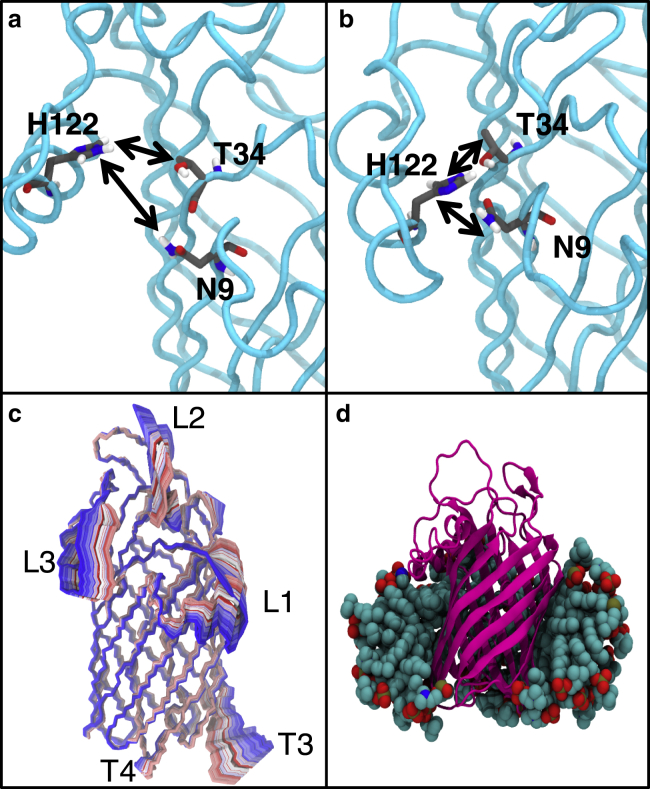
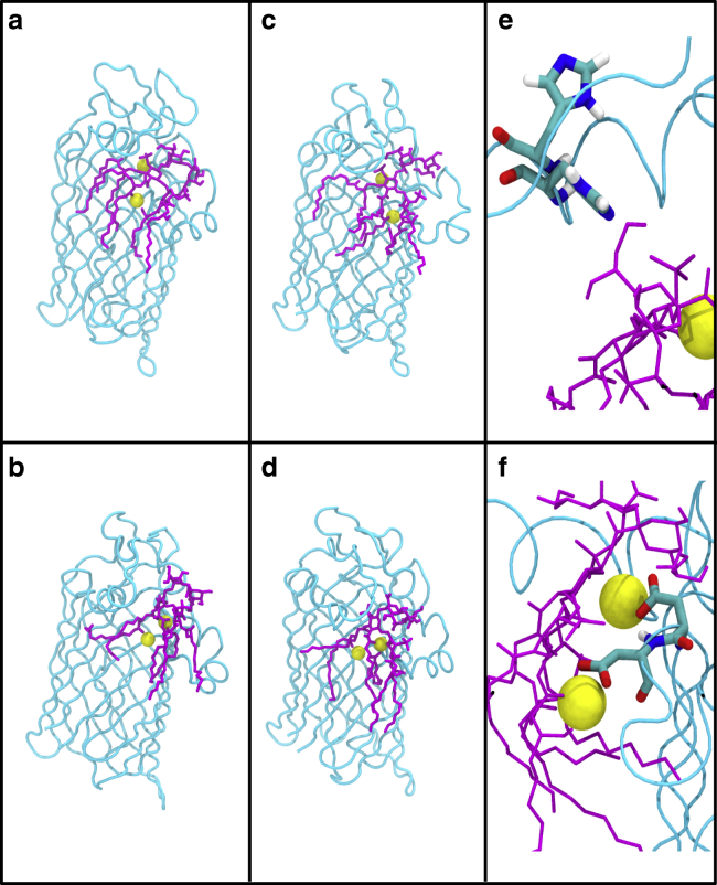
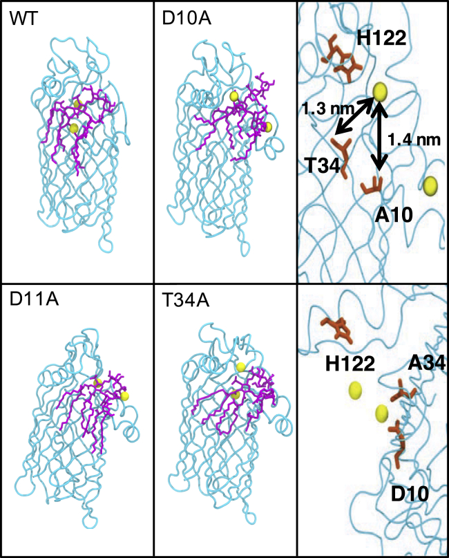
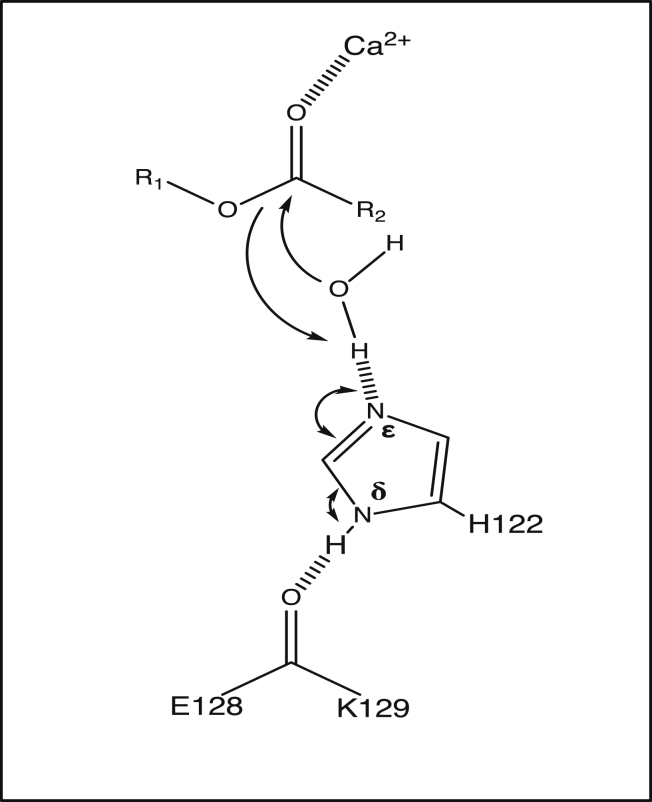
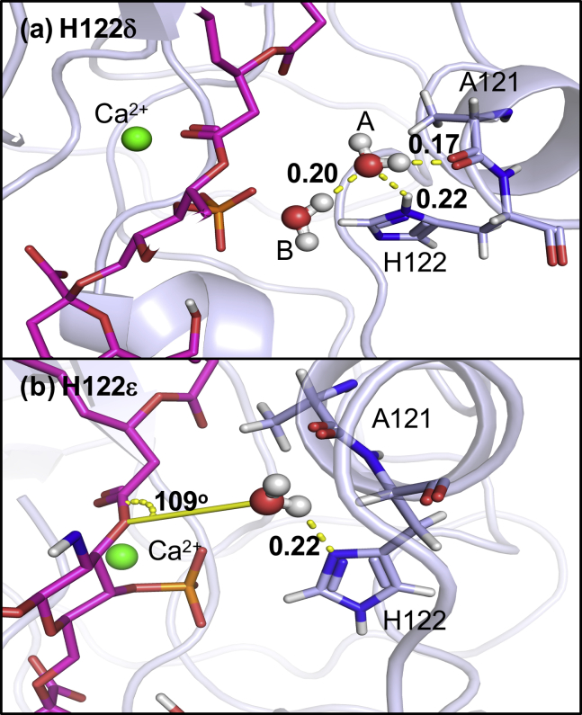
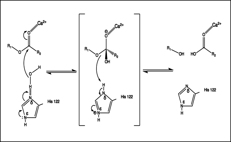
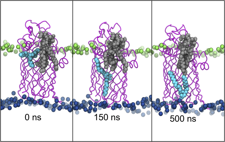
Covalent modification of outer membrane lipids of Gram-negative bacteria can impact the ability of the bacterium to develop resistance to antibiotics as well as modulating the immune response of the host. The enzyme LpxR from Salmonella typhimurium is known to deacylate lipopolysaccharide molecules of the outer membrane; however, the mechanism of action is unknown. Here, we employ molecular dynamics and Monte Carlo simulations to study the conformational dynamics and substrate binding of LpxR in representative outer membrane models as well as detergent micelles. We examine the roles of conserved residues and provide an understanding of how LpxR binds its substrate. Our simulations predict that the catalytic H122 must be Nε-protonated for a single water molecule to occupy the space between it and the scissile bond, with a free binding energy of -8.5 kcal mol-1. Furthermore, simulations of the protein within a micelle enable us to predict the structure of the putative "closed" protein. Our results highlight the need for including dynamics, a representative environment, and the consideration of multiple tautomeric and rotameric states of key residues in mechanistic studies; static structures alone do not tell the full story.


Figures

Figure 1
Figure 1
(a) Coarse-grained and (b) united-atom models of LpxR from the X-ray structure (PDB: 3FID) embedded within a model of the OM. Ra LPS headgroups are shown in lime, phospholipid headgroups are shown in blue, and lipids tails are omitted for clarity. (c) Re LPS with scissile bond are indicated and hydrogen atoms are removed for clarity. (d) A close-up of the docked Re LPS molecule (black) with residues thought to be key for catalysis through mutagenesis studies is highlighted (5). To see this figure in color, go online.
Figure 2
Figure 2
Conformational dynamics of LpxR within a detergent micelle at 323 K. (a) The position of catalytic residues in the binding pocket in the energy-minimized LpxR structure (PDB: 3FID). The protein backbone is shown in cyan, with carbon, oxygen, nitrogen, and hydrogen colored gray, red, blue, and white, respectively. (b) Closed conformation of the protein revealed by simulations in DPC micelles, with catalytic residues within hydrogen-bonding distance of each other. (c) The motion of the protein is depicted by extrapolating between the two extreme projections described by eigenvector 1 and then overlaying the conformation of the protein after every 20 ps. The image is colored on a blue, white, and red scale (blue at the start of the simulation, through white to red at the end of the simulation). (d) A representative snapshot of the protein in detergent micelle, some of the detergent molecules have been removed for clarity. To see this figure in color, go online.
Figure 3
Figure 3
Snapshots of the protein-lipid-ion complex after 300 ns from two independent simulations of the ion-biased (a and c) and ion-unbiased (b and d) simulations. The two rotameric states of H122 are shown in (e), and (f) shows coordination of cations by residues D10 and D11. The protein is colored cyan, Re LPS is magenta, and ions are yellow. The membrane, water, and other ions are omitted for clarity. To see this figure in color, go online.
Figure 4
Figure 4
Snapshots of the protein-lipid-ion complex with specific residue mutations after 300 ns, compared to the wild-type. The membrane and solvent have been omitted for clarity. The top right panel shows the cation moving away from residue 10 when it is mutated from D to A. The bottom right panel shows the cation is still near its original location when residue 34 is mutated from T to A. The color scheme is the same as Fig. 3. To see this figure in color, go online.
Figure 5
Figure 5
Catalytic mechanism including the residues E128, K129, H122, the portion of Re LPS to be cleaved, and one Ca2+ ion.
Figure 6
Figure 6
(a) Water locations from the GCMC simulation where H122 is Nδ protonated. Two water molecules found between the H122 and the ester. Hydrogen-bonding interactions are illustrated with a dashed yellow line, with length shown in nanometers. Water molecule B is forming hydrogen bonds with the H122 Hδ, water A, and A121. The water A molecule is ∼0.3 nm from the ester group throughout the simulation and is orientated with the oxygen atom pointed toward the group, which means that it is unable to hydrogen bond to the ester group and would therefore not result in hydrolysis. The water is stabilized in this orientation by hydrogen bonding to another water close by. (b) A snapshot of the Nε-protonated H122. A121, H122, and ligand are shown in stick representation, and the protein backbone is shown in light blue. The calcium ion is shown in green. The relevant water(s) in each case are shown in red and white. Part of the ligand and other GCMC water molecules have been removed from the image for clarity. To see this figure in color, go online.
Figure 7
Figure 7
The predicted mechanism of deacylation based on our molecular dynamics and GCMC simulations. The catalytic histidine, H122, is Nε protonated. To establish whether the GCMC and molecular dynamics simulations were identifying similar water sites, we defined a 1-nm3 box around H122 and the ester moiety of the ligand in the ligand-bound unbiased trajectory. Using visual molecular dynamics, we found that water molecules occupied this space for 95% of our 1-μs molecular dynamics simulation at 323 K. More than one water molecule was found when the distance between H122 and the ester moiety increased.
Figure 8
Figure 8
Movement of catalytically cleaved acyl tails (cyan) away from the protein (magenta) and the truncated Re LPS substrate (gray). To see this figure in color, go online.

Similar articles

Cited by

References

    1. Nikaido H. Molecular basis of bacterial outer membrane permeability revisited. Microbiol. Mol. Biol. Rev. 2003;67:593–656. - PMC - PubMed
    1. Kim S., Patel D.S., et al. Im W. Bilayer properties of lipid A from various Gram-negative bacteria. Biophys. J. 2016;111:1750–1760. - PMC - PubMed
    1. Hwang P.M., Kay L.E. Solution structure and dynamics of integral membrane proteins by NMR: a case study involving the enzyme PagP. Methods Enzymol. 2005;394:335–350. - PubMed
    1. Rutten L., Geurtsen J., et al. Tommassen J. Crystal structure and catalytic mechanism of the LPS 3-O-deacylase PagL from Pseudomonas aeruginosa. Proc. Natl. Acad. Sci. USA. 2006;103:7071–7076. - PMC - PubMed
    1. Rutten L., Mannie J.P., et al. Gros P. Active-site architecture and catalytic mechanism of the lipid A deacylase LpxR of Salmonella typhimurium. Proc. Natl. Acad. Sci. USA. 2009;106:1960–1964. - PMC - PubMed

Publication types

Substances

LinkOut - more resources