. 2019 Jan;25(1):23-34.
doi: 10.1261/rna.067975.118. Epub 2018 Oct 4.

Rare variants of the FMN riboswitch class in Clostridium difficile and other bacteria exhibit altered ligand specificity

Affiliations

Rare variants of the FMN riboswitch class in Clostridium difficile and other bacteria exhibit altered ligand specificity

Ruben M Atilho et al. RNA. 2019 Jan.

Abstract

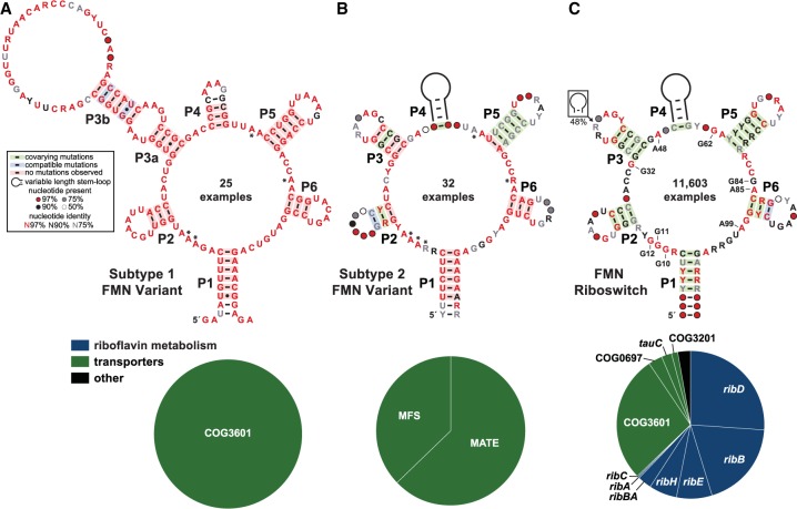
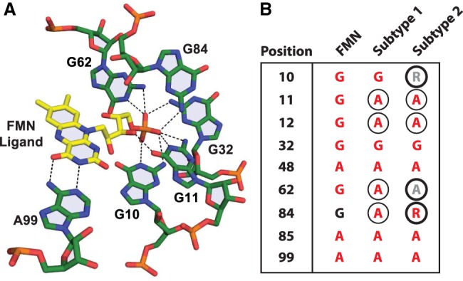
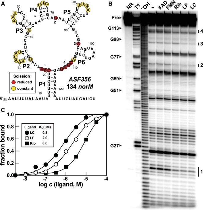
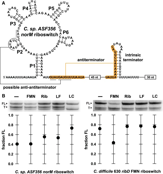
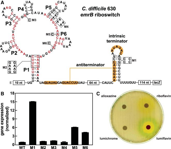
Many bacteria use flavin mononucleotide (FMN) riboswitches to control the expression of genes responsible for the biosynthesis and transport of this enzyme cofactor or its precursor, riboflavin. Rare variants of FMN riboswitches found in strains of Clostridium difficile and some other bacteria typically control the expression of proteins annotated as transporters, including multidrug efflux pumps. These RNAs no longer recognize FMN, and differ from the original riboswitch consensus sequence at nucleotide positions normally involved in binding of the ribityl and phosphate moieties of the cofactor. Representatives of one of the two variant subtypes were found to bind the FMN precursor riboflavin and the FMN degradation products lumiflavin and lumichrome. Although the biologically relevant ligand sensed by these variant FMN riboswitches remains uncertain, our findings suggest that many strains of C. difficile might use rare riboswitches to sense flavin degradation products and activate transporters for their detoxification.

Keywords: aptamer; flavin mononucleotide; lumichrome; lumiflavin; noncoding RNA; riboflavin biosynthesis.


Figures

FIGURE 1.
FIGURE 1.
Variant FMN motif RNAs differ from the FMN riboswitch consensus and its typical genetic associations. (A) Consensus sequence and secondary structure model of 25 unique examples of subtype 1 variant FMN RNAs. Asterisks identify key nucleotides that differ from FMN riboswitch consensus. The key (box) describes the annotations in the consensus models. (Bottom) Pie chart of the genes located immediately downstream from subtype 1 RNAs. (B) Consensus model and gene associations for 32 unique examples of subtype 2 RNAs. Additional annotations are as described for A. (C) Consensus model and gene associations for 11,603 unique examples of FMN riboswitch aptamers. Certain nucleotides that are most directly involved in forming the ligand binding pocket for FMN are numbered according to the crystal structure model (PDB ID code 3F2Q) published previously (Serganov et al. 2009).
FIGURE 2.
FIGURE 2.
Atomic-resolution structural model for the binding site of an FMN riboswitch from F. nucleatum (PDB ID code 3F2Q) (Serganov et al. 2009). Dashed lines represent hydrogen-bonding contacts between aptamer nucleotides (numbered as depicted in Fig. 1C) and the ligand, FMN. (B) Comparison of key equivalent nucleotide positions among FMN aptamer, subtype 1, and subtype 2 RNAs. Nucleotides are depicted according to their level of sequence conservation as described in the key to Figure 1A. Circled nucleotides are distinct from the consensus model for FMN riboswitches. Thick-lined circles identify nucleotides in the consensus for subtype 2 RNAs that differ from the consensus for subtype 1 RNAs. Nucleotide positions are depicted in A, except for positions 12, 48, and 85, which were omitted for image clarity.
FIGURE 3.
FIGURE 3.
A representative of the subtype 2 variant FMN motif RNAs recognize FMN degradation products. (A) Sequence and secondary structure model of the 134 norM RNA construct. Lowercase letters on the 5′ terminus represent guanosine nucleotides added to the DNA template to promote transcription by T7 RNA polymerase. Regions of constant and reduced spontaneous RNA cleavage upon addition of ligand are indicated by yellow and red circles, respectively. These data are derived from the in-line probing analysis depicted in B. (B) PAGE analysis of in-line probing assays with 5′ 32P-radiolabeled 134 norM RNA in the absence (−) or presence of FMN, riboflavin (Rib), lumiflavin (LF), or lumichrome (LC) at 100 µM. NR, T1, and OH designate RNAs that have undergone no reaction, that have been partially digested with RNase T1, or that have been partially digested with alkali, respectively. Bands corresponding to certain RNase T1 digestion products (scission after G residues) are labeled according to the numbering system in A. Groups of bands denoted 1 through 4 identify regions of the RNA that undergo substantial change in response to ligand addition. (C) Plot of the fraction of RNA bound to the ligand versus the logarithm of the molar concentration (c) of the ligand. Fraction bound values were estimated based on the extent of band intensity changes at region 1 from data presented in Supplemental Figure S4.
FIGURE 4.
FIGURE 4.
Subtype 2 and FMN riboswitch RNAs regulate transcription termination in response to different ligands. (A) Sequence and secondary structure model of the subtype 2 variant FMN riboswitch construct derived from the norM gene of C. sp. ASF356 used for transcription termination assays. Terminated transcripts end within the U-rich tract immediately following the intrinsic terminator stem, whereas full-length transcripts from the DNA template used for this assay include the 30 nt following the U-rich tract. (B, top) PAGE analysis of single-round transcription termination assays conducted in the absence of ligand (−), or in the presence of 1 mM FMN, riboflavin (Rib), lumiflavin (LF), or lumichrome (LC). FL and T denote full-length and terminated RNA transcripts, respectively. (Bottom) Values for the fraction of FL RNA generated are derived from the PAGE data presented.
FIGURE 5.
FIGURE 5.
In vivo assessment of gene control by a subtype 2 variant RNA from C. difficile. (A) Sequence and secondary structure of the regulatory region of the riboswitch-reporter construct made by joining the variant FMN riboswitch RNA from the emrB gene of C. difficile 630 to a lacZ gene. Red nucleotides are >97% conserved. Constructs carrying mutations to conserved nucleotides and secondary structures are designated M1 through M6. (B) Plot of the normalized β-galactosidase reporter gene expression levels of B. subtilis cells containing WT and various mutant reporter constructs in rich medium. Error bars indicate the standard deviation from three independent experiments. (C) Agar diffusion assays were conducted with LB agar plates supplemented with 100 µg mL−1 X-gal and inoculated with B. subtilis cells carrying the WT reporter construct as described in A. Filter disks were infused with 10 µL of alloxazine (50 mM), riboflavin (35 mM), lumichrome (10 mM), or lumiflavin (80 mM). Note that the yellow-orange color surrounding the riboflavin-applied disk is due to the color of this compound, which is diffusing outward.

References

    1. Baker JL, Sudarsan N, Weinberg Z, Roth A, Stockbridge RB, Breaker RR. 2012. Widespread genetic switches and toxicity resistance proteins for fluoride. Science 335: 233–235. 10.1126/science.1215063 - DOI - PMC - PubMed
    1. Barrick JE, Corbino KA, Winkler WC, Nahvi A, Mandal M, Collins J, Lee M, Roth A, Sudarsan N, Jona I, et al. 2004. New riboswitch motifs suggest an expanded scope for riboswitches in bacterial genetic control. Proc Natl Acad Sci 101: 6421–6426. 10.1073/pnas.0308014101 - DOI - PMC - PubMed
    1. Blount KF. 2013. Methods for treating or inhibiting infection by Clostridium difficile. U.S. patent appl. no. 13/576,989.
    1. Blount KF, Coish PDG, Dixon BR, Myung J, Osterman D, Wickens P, Avola S, Baboulas N, Bello A, Berman J, et al. 2012. Flavin derivatives. U.S. patent appl. no. 13/381,809.
    1. Blount KF, Megyola C, Plummer M, Osterman D, O'Connell T, Aristoff P, Quinn C, Chrusciel RA, Poel TJ, Schostarez HJ, et al. 2015. Novel riboswitch-binding flavin analog that protects mice against Clostridium difficile infection without inhibiting cecal flora. Antimicrob Agents Chemother 59: 5736–5746. 10.1128/AAC.01282-15 - DOI - PMC - PubMed

Publication types

LinkOut - more resources