Review
. 2018 Oct;210(2):357-396.
doi: 10.1534/genetics.118.300224.

Anatomy and Physiology of the Digestive Tract of Drosophila melanogaster

Affiliations
Review

Anatomy and Physiology of the Digestive Tract of Drosophila melanogaster

Irene Miguel-Aliaga et al. Genetics. 2018 Oct.

Abstract

The gastrointestinal tract has recently come to the forefront of multiple research fields. It is now recognized as a major source of signals modulating food intake, insulin secretion and energy balance. It is also a key player in immunity and, through its interaction with microbiota, can shape our physiology and behavior in complex and sometimes unexpected ways. The insect intestine had remained, by comparison, relatively unexplored until the identification of adult somatic stem cells in the Drosophila intestine over a decade ago. Since then, a growing scientific community has exploited the genetic amenability of this insect organ in powerful and creative ways. By doing so, we have shed light on a broad range of biological questions revolving around stem cells and their niches, interorgan signaling and immunity. Despite their relatively recent discovery, some of the mechanisms active in the intestine of flies have already been shown to be more widely applicable to other gastrointestinal systems, and may therefore become relevant in the context of human pathologies such as gastrointestinal cancers, aging, or obesity. This review summarizes our current knowledge of both the formation and function of the Drosophila melanogaster digestive tract, with a major focus on its main digestive/absorptive portion: the strikingly adaptable adult midgut.

Keywords: Drosophila; FlyBook; absorption; aging; digestion; enteric nervous system; enteroendocrine; immunity; intestine; metals; microbiota; midgut; stem cells.


Figures

Figure 1
Figure 1
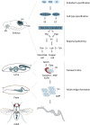
Developmental transitions and key factors in intestinal cell fate decisions. See section Embryonic and larval development for details.
Figure 2
Figure 2
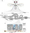
The adult intestine and its cell types. (A) The digestive tract is highlighted in gray inside an adult fly. (B) Main anatomical features of the adult digestive tract. (C) General cellular composition of the digestive tract. See section The adult gut and its cell types: genetic and anatomical compartmentalization for details.
Figure 3
Figure 3
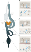
Regional differences in ISC proliferation. (A) General mode of midgut ISC proliferation. See main text for details in The adult gut and its cell types: genetic and anatomical compartmentalization. Alternative modes of ISC proliferation - stress induced rather than constitutive - are found in two specific intestinal regions: the copper cell region (B) and the hindgut (C). See main text in The adult gut and its cell types: genetic and anatomical compartmentalization for details. CC, copper cell; GB, gastroblast, GSSCs, gastric stem cells; IC, interstitial cell.
Figure 4
Figure 4
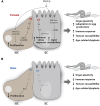
Sex and reproductive differences in ISCs and ECs. (A) Contributions of the intrinsic sex differentiation pathway and the mating-triggered rise in circulating JH to ISC and EC homeostasis in females. (B) Contributions of the intrinsic sex differentiation pathway to ISC and EC homeostasis in males. See section Sex differences and reproductive plasticity for details. This figure was inspired by Portman and Biteau (2016).
Figure 5
Figure 5
Innervation of the adult intestine. (A) Enteric innervation of the adult digestive tract. Neurons located in the brain and enteric ganglia contribute to the innervation of anterior portions (see B and Gut-innervating neurons section for details). Central neurons with cell bodies in the posterior segments of the abdominal ganglion of the ventral nerve cord (VNC) send axons in the hindgut nerves toward the pylorus, which extend anterior along the posterior portion of the midgut. The hindgut nerves also branch to innervate the rectal ampulla and rectum. (B) Stomatogastric nervous system ganglia and nerves contribute to enteric innervation of the larval esophagus and anterior midgut (left) and adult esophagus, crop, and anterior midgut (right). In adults, the larval proventricular and hypocerebral ganglia have fused. The fused hypocerebral ganglion is adjacent to the corpus allatum and corpora cardiaca, which have moved posteriorly from their larval head position to the main body of the adult fly.

Similar articles

Cited by

References

    1. Abraham I., Doane W. W., 1978. Genetic regulation of tissue-specific expression of amylase structural genes in Drosophila melanogaster. Proc. Natl. Acad. Sci. USA 75: 4446–4450. 10.1073/pnas.75.9.4446 - DOI - PMC - PubMed
    1. Abu F., Wang J. G., Oh Y., Deng J., Neubert T. A., et al. , 2018. Communicating the nutritional value of sugar in Drosophila. Proc. Natl. Acad. Sci. USA 115: E2829–E2838. 10.1073/pnas.1719827115 - DOI - PMC - PubMed
    1. Alfa R. W., Park S., Skelly K. R., Poffenberger G., Jain N., et al. , 2015. Suppression of insulin production and secretion by a decretin hormone. Cell Metab. 21: 323–334 (corrigenda: Cell Metab. 27: 479 (2018)) 10.1016/j.cmet.2015.01.006 - DOI - PMC - PubMed
    1. Allan A. K., Du J., Davies S. A., Dow J. A., 2005. Genome-wide survey of V-ATPase genes in Drosophila reveals a conserved renal phenotype for lethal alleles. Physiol. Genomics 22: 128–138. 10.1152/physiolgenomics.00233.2004 - DOI - PubMed
    1. Amcheslavsky A., Jiang J., Ip Y. T., 2009. Tissue damage-induced intestinal stem cell division in Drosophila. Cell Stem Cell 4: 49–61. 10.1016/j.stem.2008.10.016 - DOI - PMC - PubMed

Publication types

MeSH terms