. 2018 Nov 16;362(6416):808-813.
doi: 10.1126/science.aat6141. Epub 2018 Oct 4.

Reversible self-assembly of superstructured networks

Affiliations

Reversible self-assembly of superstructured networks

Ronit Freeman et al. Science. .

Abstract

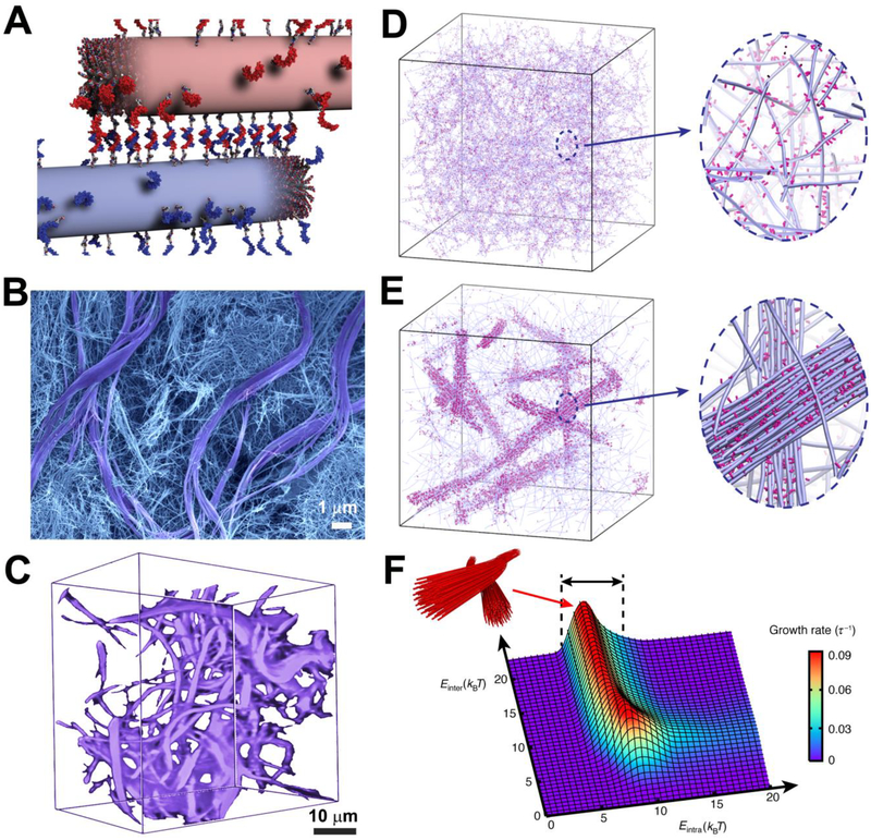
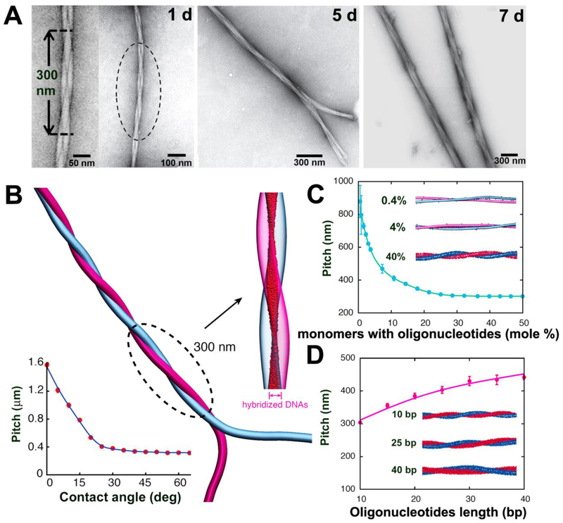
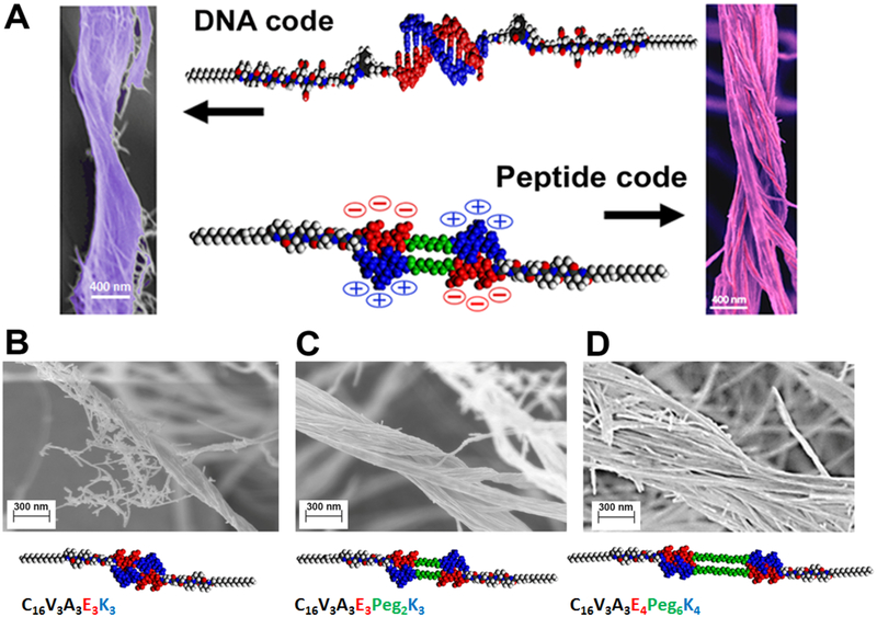
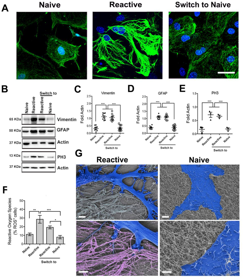
Soft structures in nature, such as protein assemblies, can organize reversibly into functional and often hierarchical architectures through noncovalent interactions. Molecularly encoding this dynamic capability in synthetic materials has remained an elusive goal. We report on hydrogels of peptide-DNA conjugates and peptides that organize into superstructures of intertwined filaments that disassemble upon the addition of molecules or changes in charge density. Experiments and simulations demonstrate that this response requires large-scale spatial redistribution of molecules directed by strong noncovalent interactions among them. Simulations also suggest that the chemically reversible structures can only occur within a limited range of supramolecular cohesive energies. Storage moduli of the hydrogels change reversibly as superstructures form and disappear, as does the phenotype of neural cells in contact with these materials.


Conflict of interest statement

Competing interests: None declared

Figures

Figure 1.
Figure 1.. Dynamics in DNA-peptide amphiphiles drives the formation of hierarchical structures
(A) Illustration of peptide amphiphile fibers cross-linked by DNA hybridization; fibers are shown in their initial state prior to monomer exchange. (B) SEM micrograph of the hydrogel formed upon DNA cross-linking showing two populations within the gel, consisting of twisted bundles (diameter ~1–3 μm) and single fibers (diameters between 10 and 15 nm). (C) Confocal reconstruction image of a section of the gel containing DNA monomers modified with the fluorescent dye Cy3. Bundles are shown in purple. (D) Simulation snapshots showing a homogeneous hydrogel when molecular exchange of DNA monomers between PA fibers is prohibited. Magnified view shows individual fibers (blue) with a stochastic distribution of DNA monomers (pink) along the fibers. (E) Simulation snapshots showing the emergence of bundles of fibers when molecular exchange is allowed. Magnified view shows bundle of fibers (blue) enriched with DNA (pink) in a matrix of individual fibers depleted of DNA monomers. (F) Bundle growth rate as a function of intra- and inter-fiber energies (Eintra, Einter). Bundles form within the energy range 5 kBT < Eintra < 10 kBT (black arrows).
Figure 2.
Figure 2.. Programming the growth of intertwined bundles of fibers
(A) TEM images after mixing complementary DNA- and PNA-terminated peptide amphiphiles show the time-dependent evolution of twisted bundles over 24 hours, 5 days, and 7 days. (B) Simulation snapshot of two intertwined complementary fibers. The intertwining pitch saturates for most initial contact angles (bottom left). Hybridized DNA-PNA pairs between the two fibers (magnified view) form a twisted ribbon pattern. (C) Dependence of the pitch on the fraction of monomers with oligonucleotides. Simulation snapshots shown for systems with 0.4%, 4%, and 40% oligonucleotides-modified monomers. (D) Dependence of the pitch on the oligonucleotides length. Simulation snapshots shown for duplexes with 10, 25, and 40 DNA-PNA base pairs (bp).
Figure 3.
Figure 3.. Programming hierarchical structures with a peptide code
(A) Molecular graphics representation of the complementary interactions between the DNA (top) and DNA-mimetic peptide amphiphiles (bottom), and the corresponding morphologies of bundled fibers observed in both systems by scanning electron microscopy. (B-D) Scanning electron micrographs of bundled and twisted fiber morphologies of varying diameters: (B) 140.5 ± 15 nm; (C) 332 ± 37 nm, and (D) 905 ± 190 nm, and the corresponding dimer molecular graphics and chemical sequences of the DNA-mimetic peptide amphiphiles that form the superstructures (EG refers to ethylene oxide and C16 is the number of carbons in the aliphatic terminus of the amphiphiles). Quantification of bundle diameters utilized a minimum of 15 randomly selected images (taken from three independent batches) for each system.
Figure 4.
Figure 4.. Modulating the phenotype of astrocytes on reversible hierarchical ECM mimetic
(A) Confocal microscopy images of astrocytes plated on individual fibers (left), on bundled fibers (center), and after switching from bundles to individual fibers (right). Staining for GFAP (green) and cell nuclei (DAPI, blue) reveals cells with naive morphology on substrates of individual fibers and reactive morphology on substrates of bundled fibers. Scale bar: 50 μm, pertaining to all images. (B) Western blot analysis of protein expression (related to cytoskeleton and cell proliferation) in astrocytes on indicated substrates. (C-E) Relative expression of proteins derived from western blots in B. All values were normalized to Actin expression; three experiments were analyzed. (*p<0.05; **p<0.01; ****p<0.0001, LSD test). (F) Reactive oxygen species (ROS) quantification on the different substrates relative to cell number. (*p<0.05; **p<0.01; ****p<0.0001). (G) SEM micrographs of a reactive cell on bundled fibers and a naïve cell on individual fibers. Cells are falsely colored in blue. The magnified view (lower images) shows the cell-substrate interaction. Bundles are falsely colored in pink. Scale bars: 5 μm (upper images), 2 μm (lower images).

References

    1. Whitesides GM, Grzybowski B, Self-assembly at all scales. Science 295, 2418–2421 (2002). - PubMed
    1. Zhang S, Fabrication of novel biomaterials through molecular self-assembly. Nature biotechnology 21, 1171–1178 (2003). - PubMed
    1. Needleman D, Dogic Z, Active matter at the interface between materials science and cell biology. Nature Reviews Materials 2, 17048 (2017).
    1. Ridley AJ, Hall A, The small GTP-binding protein rho regulates the assembly of focal adhesions and actin stress fibers in response to growth factors. Cell 70, 389–399 (1992). - PubMed
    1. Dos Remedios C et al., Actin binding proteins: regulation of cytoskeletal microfilaments. Physiological reviews 83, 433–473 (2003). - PubMed

Publication types