. 2018 Dec 21;293(51):19532-19543.
doi: 10.1074/jbc.RA118.004909. Epub 2018 Oct 4.

Simulations of the regulatory ACT domain of human phenylalanine hydroxylase (PAH) unveil its mechanism of phenylalanine binding

Affiliations

Simulations of the regulatory ACT domain of human phenylalanine hydroxylase (PAH) unveil its mechanism of phenylalanine binding

Yunhui Ge et al. J Biol Chem. .

Abstract

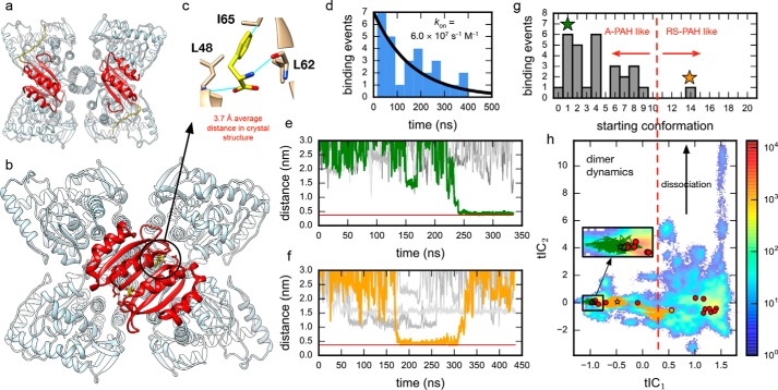
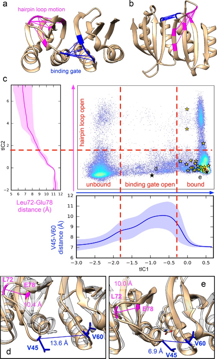
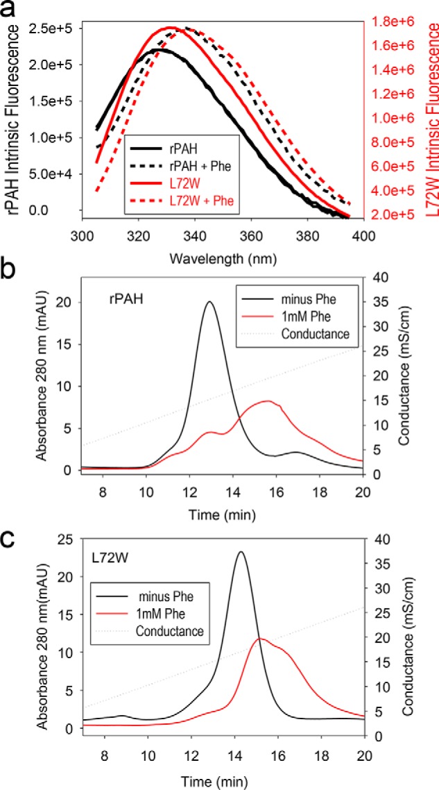
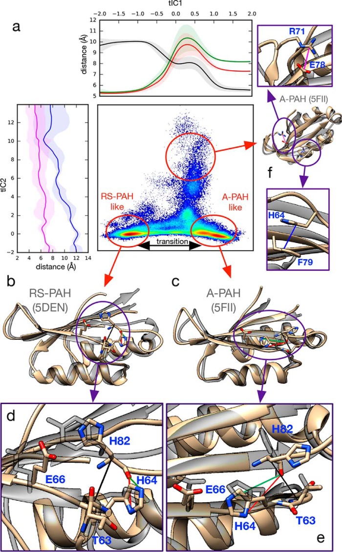
Phenylalanine hydroxylase (PAH) regulates phenylalanine (Phe) levels in mammals to prevent neurotoxicity resulting from high Phe concentrations as observed in genetic disorders leading to hyperphenylalaninemia and phenylketonuria. PAH senses elevated Phe concentrations by transient allosteric Phe binding to a protein-protein interface between ACT domains of different subunits in a PAH tetramer. This interface is present in an activated PAH (A-PAH) tetramer and absent in a resting-state PAH (RS-PAH) tetramer. To investigate this allosteric sensing mechanism, here we used the GROMACS molecular dynamics simulation suite on the Folding@home computing platform to perform extensive molecular simulations and Markov state model (MSM) analysis of Phe binding to ACT domain dimers. These simulations strongly implicated a conformational selection mechanism for Phe association with ACT domain dimers and revealed protein motions that act as a gating mechanism for Phe binding. The MSMs also illuminate a highly mobile hairpin loop, consistent with experimental findings also presented here that the PAH variant L72W does not shift the PAH structural equilibrium toward the activated state. Finally, simulations of ACT domain monomers are presented, in which spontaneous transitions between resting-state and activated conformations are observed, also consistent with a mechanism of conformational selection. These mechanistic details provide detailed insight into the regulation of PAH activation and provide testable hypotheses for the development of new allosteric effectors to correct structural and functional defects in PAH.

Keywords: Markov state models; allosteric regulation; binding pathways; conformational change; conformational selection; kinetics; ligand binding kinetics; ligand-binding protein; molecular dynamics; molecular simulation; phenylalanine hydroxylase; phenylketonuria.


Conflict of interest statement

The authors declare that they have no conflicts of interest with the contents of this article

Figures

Figure 1.
Figure 1.
Ab initio binding simulations implicate a conformational selection mechanism for Phe association with ACT domain dimers. a, the crystal structure of full-length tetrameric mammalian (rat) RS-PAH (PDB code 5DEN) with two ACT domains shown in red. The other two ACT domains, uncolored, are in the back. b, a model of A-PAH (human) prepared using the crystal structure of the Phe-bound ACT domain dimer (PDB code 5FII). Again, two ACT domains are colored red, and the two uncolored ACT domains are in the back. c, average distances (Phe-C)–(Leu48-N), (Phe-Cζ)–(Ile65-Cβ), and (Phe-N)–(Leu62-O) were used to monitor productive binding events using a threshold of 0.375 nm. d, binding time distribution for 29 observed binding events shown with a Bayesian estimate of the binding rate. e and f, example traces for Phe binding trajectories (orange and green) shown with example nonbinding trajectories (gray). The red line shows the average distance in the crystal structure (PDB code 5FII). g, histogram of binding events for initial conformations of the dimer suggests that a crystal-like dimer pose is necessary for productive binding. Colored stars mark the starting dimer conformations of the traces shown in e and f. Only one of the 29 binding trajectories starts from a dimer pose close to the homology model (orange); this same trajectory also contains the only unbinding event observed (see f). h, dimer trajectory data projected to the 2D tICA landscape. Circles mark the initial dimer poses with colored traces and binding events (stars) shown for the trajectories in e and f.
Figure 2.
Figure 2.
Simulated binding pathways reveal a ligand gating mechanism. a and b, the slowest dynamics (along tIC1) corresponds to a binding gate motion coupled to Phe association, whereas the next-slowest dynamics corresponds to a hairpin loop motion. The interresidue distances that change greatly during these motions are shown in blue and pink, respectively. c, trajectory data are shown projected to the 2D tICA landscape along with average atomic distances (Val45-Cα)–(Val60-Cα) (blue) and (Leu72-Cβ)–(Glu78-Cα) (magenta) as a function tIC1 and tIC2, respectively. Yellow stars denote Phe binding events. Black stars show the locations of snapshots shown in d and e. Representative snapshots (tan) are shown of an open binding gate conformation (b) and the Phe-bound structure after binding (e). For comparison, each are superposed with the crystal structure of Phe-bound ACT domain dimer (transparent gray) (PDB code 5FII).
Figure 3.
Figure 3.
Evaluation of the RS-PAH ⇔ A-PAH equilibrium for rPAH and L72W. a, intrinsic protein fluorescence for rPAH (black) and L72W (red) in the absence (solid lines) and presence (dashed lines) of 1 mm Phe. b, ion exchange behavior of rPAH at 0 (black) and 1 mm (red) Phe. c, ion exchange behavior of L72W at 0 (black) and 1 mm (red) Phe. mAU, milli-absorbance units; mS, millisiemens.
Figure 4.
Figure 4.
Simulations of the ACT domain monomer reveal spontaneous transitions between RS-PAH–like and A-PAH–like conformations in the absence of Phe. a, monomer trajectory data projected to the 2D tICA landscape shows that RS-PAH–like (b) and A-PAH–like (c) conformations are significantly populated metastable states. Atomic distances are shown to highlight structural changes: along tIC1, (His64-Nδ1)–(His82-O) (red), (His64-Nϵ2)–(His82-O) (green), and (Thr63-Cα)–(His82-Cα) (black); along tIC2, (Arg78-Cζ)–(Glu78-Cδ) (pink) and (His64-Cγ)–(Phe79-Cγ) (blue). Ribbon structures (tan) show selected conformations from the simulation trajectory data superposed with ACT domain crystal structures (transparent gray) representative of the RS-PAH state (PDB code 5DEN) and A-PAH state (PDB code 5FII). Key structural details for RS-PAH–like (d) and A-PAH–like (e) states are shown. f, an off-pathway conformational state sampled by the simulations.

References

    1. Aravind L., and Koonin E. V. (1999) Gleaning non-trivial structural, functional and evolutionary information about proteins by iterative database searches. J. Mol. Biol. 287, 1023–1040 10.1006/jmbi.1999.2653 - DOI - PubMed
    1. Jaffe E. K. (2017) New protein structures provide an updated understanding of phenylketonuria. Mol. Genet. Metab. 121, 289–296 10.1016/j.ymgme.2017.06.005 - DOI - PMC - PubMed
    1. Jaffe E. K., Stith L., Lawrence S. H., Andrake M., and Dunbrack R. L. Jr. (2013) A new model for allosteric regulation of phenylalanine hydroxylase: implications for disease and therapeutics. Arch. Biochem. Biophys. 530, 73–82 10.1016/j.abb.2012.12.017 - DOI - PMC - PubMed
    1. Patel D., Kopec J., Fitzpatrick F., McCorvie T. J., and Yue W. W. (2016) Structural basis for ligand-dependent dimerization of phenylalanine hydroxylase regulatory domain. Sci. Rep. 6, 23748 10.1038/srep23748 - DOI - PMC - PubMed
    1. Kobe B., Jennings I. G., House C. M., Michell B. J., Goodwill K. E., Santarsiero B. D., Stevens R. C., Cotton R. G., and Kemp B. E. (1999) Structural basis of autoregulation of phenylalanine hydroxylase. Nat. Struct. Biol. 6, 442–448 10.1038/8247 - DOI - PubMed

Publication types