. 2018 Dec 7;293(49):19137-19147.
doi: 10.1074/jbc.RA118.005430. Epub 2018 Oct 4.

The C terminus of the bacterial multidrug transporter EmrE couples drug binding to proton release

Affiliations

The C terminus of the bacterial multidrug transporter EmrE couples drug binding to proton release

Nathan E Thomas et al. J Biol Chem. .

Abstract

Ion-coupled transporters must regulate access of ions and substrates into and out of the binding site to actively transport substrates and minimize dissipative leak of ions. Within the single-site alternating access model, competitive substrate binding forms the foundation of ion-coupled antiport. Strict competition between substrates leads to stoichiometric antiport without slippage. However, recent NMR studies of the bacterial multidrug transporter EmrE have demonstrated that this multidrug transporter can simultaneously bind drug and proton, which will affect the transport stoichiometry and efficiency of coupled antiport. Here, we investigated the nature of substrate competition in EmrE using multiple methods to measure proton release upon the addition of saturating concentrations of drug as a function of pH. The resulting proton-release profile confirmed simultaneous binding of drug and proton, but suggested that a residue outside EmrE's Glu-14 binding site may release protons upon drug binding. Using NMR-monitored pH titrations, we trace this drug-induced deprotonation event to His-110, EmrE's C-terminal residue. Further NMR experiments disclosed that the C-terminal tail is strongly coupled to EmrE's drug-binding domain. Consideration of our results alongside those from previous studies of EmrE suggests that this conserved tail participates in secondary gating of EmrE-mediated proton/drug transport, occluding the binding pocket of fully protonated EmrE in the absence of drug to prevent dissipative proton transport.

Keywords: EmrE; antibiotics; antiporter; efflux; gating; membrane transport; multidrug transporter; nuclear magnetic resonance (NMR); proton motive forces; proton transport; structure-function.


Conflict of interest statement

The authors declare that they have no conflicts of interest with the contents of this article

Figures

Figure 1.
Figure 1.
Two competing models for antiport. In pure exchange (left), strict competition for the single site between drug and two protons, along with prohibition of alternating access in the absence of substrate or ion, leads to tightly coupled stoichiometric antiport of 2H+/drug. In free exchange, competition remains, but no restrictions are placed on binding or alternating access, allowing both 1H+/drug (orange) and 2H+/drug (red) antiport.
Figure 2.
Figure 2.
Structure of TPP+-bound EmrE. The crystal structure (Protein Data Bank code 3B5D) (23) with monomer A in magenta, monomer B in blue, and TPP+ in orange is shown. The Cα of Glu-14 is shown as a green sphere. The final residue in the structure, Ser-105A, is indicated by a red dot. The location of the C-terminal tail is unknown, but it could interact with positively charged residues in the monomer B loops (dotted line) or fold into the binding pocket itself (dashed line).
Figure 3.
Figure 3.
Glu-14 alone cannot account for drug-induced proton release. A, proton release per EmrE dimer upon addition of saturating concentration of TPP+ as predicted by pure exchange (black line) or free exchange models of transport at 25 (blue line) or 45 °C (red line), assuming Glu-14 is the only residue involved. Experimental proton release, whether measured directly at 25 °C (blue circles) or by ITC at 45 °C (red circles), is more consistent with the free exchange model at low pH where the two models are most distinct. However, more protons are released upon TPP+ binding near neutral pH than would be predicted by the Glu-14–only free exchange model. Error bars indicate the S.E. of the fit of proton release at each pH value. B, fit of proton release measured directly at 25 °C and pH 6 by pH electrode as a function of EmrE concentration. The slope indicates the drug-induced proton release per dimer (sample data shown in D), and the error bars indicate the S.D. of standard HCl aliquots. C, fit of enthalpy of TPP+ binding as a function of buffer ionization enthalpy at 45 °C and pH 7.5. The negative slope represents the proton release per monomer (sample data shown in E). Error bars indicate the S.E. in the fit of ΔHbinding. D, representative trace of TPP+-induced proton release at pH 6. At the indicated time point, 5 μmol of TPP+ was added to a solution containing 7.5 nmol of EmrE dimer, causing a release in protons seen by the drop in pH. This pH drop was converted to nmol of H+ by the subsequent addition of known quantities of NaOH and HCl to the solution. Additional aliquots of HCl and NaOH were added to improve quantitation (data not shown). E, overlay of representative ITC binding curves for TPP+ at 45 °C and pH 7.5 in either potassium phosphate (gray), BES (dark blue), MOPS (beige), imidazole (brown), or Tris (light blue) buffer (see Table S1 for complete ITC data).
Figure 4.
Figure 4.
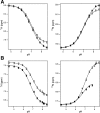
NMR-monitored pH titration of His-110 side chain. A, His-110 peaks for each monomer in HMBC NMR spectra of drug-free EmrE in isotropic bicelles are clearly resolved at 25 °C. Three or four peaks are observed for each imidazole ring as explained in Fig. S5. As pH changes, the peak positions titrate along a line, consistent with a single protonation event. The peak positions at low pH fall along this line for both monomers, indicating that the peak overlap at low pH is due to similar chemical environments for the two residues, not fast exchange. Thus, the pKa for each monomer can be determined separately. Representative peaks are shown (see Fig. S6 for full titration). B, during alternating access conformational exchange, EmrE's antiparallel protomers swap conformation. Monomer A is defined as the protomer in which the N and C termini (His-110A) are facing the same side of the membrane as the binding pocket regardless of whether EmrE is open-up or open-down.
Figure 5.
Figure 5.
Determination of the His-110 side chain pKa. A, the Hδ2 and Nϵ2 chemical shifts of His-110A (filled) and His-110B (open) from the HMBC pH titration of drug-free EmrE are fit to determine the pKa. Hδ2 and Nϵ2 data were simultaneously fit to a single pKa for each monomer individually. B, fit of His-110A (filled) and His-110B (open) from the HMBC pH titration of TPP+-bound EmrE. Proton and nitrogen were fit together, but due to line broadening at high pH, there are no nitrogen data above pH 7.77 for His-110A. A summary of pKa values can be found in Table 1. Error bars indicate the peak width at half the maximum intensity.
Figure 6.
Figure 6.
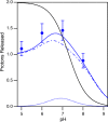
His-110 contributes to drug-induced proton release at 25 °C. Predicted proton release due to the drug-induced pKa shift of Glu-14 (dashed line) or His-110 (dotted line) measured by NMR is shown. Including both Glu-14 and His-110 in the proton-release model (solid line) leads to increased proton release near neutral pH and gives a better fit of the data compared with an Glu-14–only model (dashed line). Error bars indicate the S.E. of the fit of proton release at each pH value.
Figure 7.
Figure 7.
His-110 is protected from solvent at low pH. TROSY-HSQC NMR spectra of TPP+-bound EmrE in the absence (top) and presence (bottom) of the paramagnetic ion Mn2+ (0.5 mm) are shown. At low pH, the His-110 signal is not fully relaxed by Mn2+, indicating that it is at least partially protected from water under these conditions.
Figure 8.
Figure 8.
His-110 exists in multiple states when TPP+ is bound. TROSY-HSQC spectra of TPP+-bound EmrE selectively labeled with [13C,15N]histidine reveal at least two additional histidine peaks at both low and high pH. These could correspond to two additional states of His-110A or one additional state each of both His-110A and His-110B.

References

    1. Forrest L. R., Krämer R., and Ziegler C. (2011) The structural basis of secondary active transport mechanisms. Biochim. Biophys. Acta 1807, 167–188 10.1016/j.bbabio.2010.10.014 - DOI - PubMed
    1. Boudker O., and Verdon G. (2010) Structural perspectives on secondary active transporters. Trends Pharmacol. Sci. 31, 418–426 10.1016/j.tips.2010.06.004 - DOI - PMC - PubMed
    1. Yerushalmi H., Lebendiker M., and Schuldiner S. (1995) EmrE, an Escherichia coli 12-kDa multidrug transporter, exchanges toxic cations and H+ and is soluble in organic solvents. J. Biol. Chem. 270, 6856–6863 10.1074/jbc.270.12.6856 - DOI - PubMed
    1. Schuldiner S. (2009) EmrE, a model for studying evolution and mechanism of ion-coupled transporters. Biochim. Biophys. Acta 1794, 748–762 10.1016/j.bbapap.2008.12.018 - DOI - PubMed
    1. Muth T. R., and Schuldiner S. (2000) A membrane-embedded glutamate is required for ligand binding to the multidrug transporter EmrE. EMBO J. 19, 234–240 10.1093/emboj/19.2.234 - DOI - PMC - PubMed

Publication types

Associated data

LinkOut - more resources