. 2018 Nov 23;293(47):18318-18327.
doi: 10.1074/jbc.RA118.004213. Epub 2018 Oct 4.

Scrambling of natural and fluorescently tagged phosphatidylinositol by reconstituted G protein-coupled receptor and TMEM16 scramblases

Affiliations

Scrambling of natural and fluorescently tagged phosphatidylinositol by reconstituted G protein-coupled receptor and TMEM16 scramblases

Lei Wang et al. J Biol Chem. .

Abstract

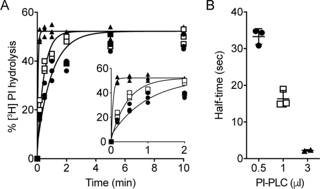
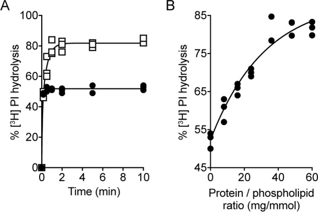
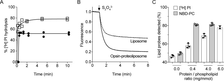
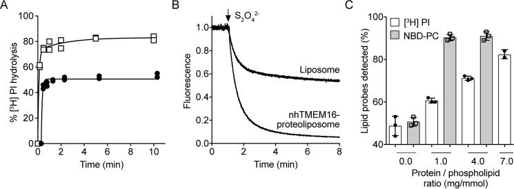
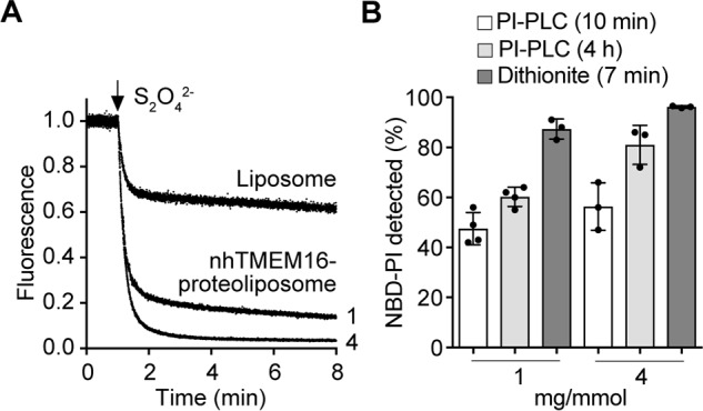
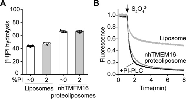
Members of the G protein-coupled receptor and TMEM16 (transmembrane protein 16) protein families are phospholipid scramblases that facilitate rapid, bidirectional movement of phospholipids across a membrane bilayer in an ATP-independent manner. On reconstitution into large unilamellar vesicles, these proteins scramble more than 10,000 lipids/protein/s as measured with co-reconstituted fluorescent nitrobenzoxadiazole (NBD)-labeled phospholipids. Although NBD-labeled phospholipids are ubiquitously used as reporters of scramblase activity, it remains unclear whether the NBD modification influences the quantitative outcomes of the scramblase assay. We now report a refined biochemical approach for measuring the activity of scramblase proteins with radiolabeled natural phosphatidylinositol ([3H]PI) and exploiting the hydrolytic activity of bacterial PI-specific phospholipase C (PI-PLC) to detect the transbilayer movement of PI. PI-PLC rapidly hydrolyzed 50% of [3H]PI in large symmetric, unilamellar liposomes, corresponding to the lipid pool in the outer leaflet. On reconstitution of a crude preparation of yeast endoplasmic reticulum scramblase, purified bovine opsin, or purified Nectria haematococca TMEM16, the extent of [3H]PI hydrolysis increased, indicating that [3H]PI from the inner leaflet had been scrambled to the outer leaflet. Using transphosphatidylation, we synthesized acyl-NBD-PI and used it to compare our PI-PLC-based assay with conventional fluorescence-based methods. Our results revealed quantitative differences between the two assays that we attribute to the specific features of the assays themselves rather than to the nature of the phospholipid. In summary, we have developed an assay that measures scrambling of a chemically unmodified phospholipid by a reconstituted scramblase.

Keywords: G protein–coupled receptor (GPCR); Phospholipase C; TMEM16; glycerophospholipid; liposome; membrane transport; phosphatidylinositol; scramblase.


Conflict of interest statement

The authors declare that they have no conflicts of interest with the contents of this article

Figures

Figure 1.
Figure 1.
Schematic of PI-PLC assay. Incubation of unilamellar liposomes composed of egg PC and egg PA (9:1, molar ratio; green symbols) and trace amounts of [3H]PI (red symbols) with PI-PLC results in hydrolysis of [3H]PI (to inositol cyclic phosphate and diacylglycerol (DAG)) in the outer leaflet of the bilayer, whereas [3H]PI in the inner leaflet is protected from the enzyme. Because [3H]PI distributes equally between the two leaflets during preparation of liposomes, ∼50% is expected to be hydrolyzed by PI-PLC. In unilamellar proteoliposomes reconstituted with scramblase protein(s), [3H]PI from the inner leaflet is translocated to the outer leaflet, where it becomes accessible to PI-PLC. The extent of [3H]PI hydrolysis in scramblase-containing proteoliposomes is expected to reach 100% on a relatively short time scale.
Figure 2.
Figure 2.
PI-PLC–mediated hydrolysis of [3H]PI in liposomes. A, liposomes composed of egg PC, egg PA (9:1, molar ratio), and trace amounts of [3H]PI were treated with different amounts of PI-PLC at room temperature. At the indicated times, aliquots of the suspensions were removed, and lipids were extracted using a two-phase extraction system. Radioactivity in the upper aqueous and lower organic phases, containing 1,2-cyclic [3H]inositol phosphate/[3H]inositol 1-phosphate and ([3H]PI), respectively, was used to calculate the fraction of [3H]PI hydrolyzed by PI-PLC. The data points are from three independent experiments done in the presence of 0.5 μl (filled circles), 1.0 μl (open squares), or 3.0 μl (filled triangles) PI-PLC. In control incubations in the absence of PI-PLC, [3H]PI hydrolysis was <3%. For better visibility, the inset shows the curves during the first 2 min of incubation. The data were fitted to a one-phase exponential function. B, half-times of [3H]PI hydrolysis in the presence of different amounts of PI-PLC calculated from the curves shown in A. The data are mean values ± standard deviations from three independent experiments.
Figure 3.
Figure 3.
PI-PLC–mediated hydrolysis of [3H]PI in proteoliposomes reconstituted with yeast Triton extract. Preformed liposomes composed of egg PC and egg PA (9:1, molar ratio) were reconstituted with trace amounts of [3H]PI and a Triton extract from S. cerevisiae containing phospholipid scrambling activity. A, proteoliposomes (open squares) and control liposomes (mock reconstituted; filled circles) were treated with 3 μl of PI-PLC at room temperature, and the extent of [3H]PI hydrolysis was determined as described in Fig. 2A. The data points are from three independent experiments. The time course for [3H]PI hydrolysis in proteoliposomes was fitted to a double-exponential function. To display all data points, the graph of mock-treated liposomes was shifted by +0.3 min on the x axis. B, dependence of [3H]PI hydrolysis on the protein to phospholipid ratio. Maximal [3H]PI hydrolysis after 10 min of PI-PLC treatment was determined in proteoliposomes reconstituted with different amounts of yeast Triton extract and plotted against the protein to phospholipid ratio. The data points are from three independent experiments and fitted into a one-phase exponential function.
Figure 4.
Figure 4.
Opsin-mediated scrambling of [3H]PI and NBD-PC. A, proteoliposomes composed of egg PC and egg PA (9:1, molar ratio) were reconstituted with [3H]PI and purified opsin using protein to phospholipid ratios of 0 (filled circles) or ∼6 (open squares) mg/mmol. The extent of PI-PLC–mediated hydrolysis of [3H]PI was determined at each time point as described in Fig. 2A. The data are from two independent experiments. The time course for [3H]PI hydrolysis in proteoliposomes was fitted to a double-exponential function. To display all data points, the graphs of opsin-liposomes at ∼3 mg/mmol and mock-treated liposomes were shifted by +0.3 and +0.6 min, respectively, on the x axis. B, liposomes composed of egg PC and egg PA (9:1 molar ratio) were reconstituted with trace amounts of NBD-PC in the absence or presence of purified opsin using a protein to phospholipid ratio of ∼0.4 mg/mmol. Dithionite (to reduce the fluorescent signal of NBD-PC) was added after 60 s, and fluorescence was recorded at room temperature with constant stirring. The traces are representative of three independent experiments and reflect opsin liposomes (solid line) and mock-reconstituted liposomes (dashed line). C, proteoliposomes composed of egg PC and egg PA (9:1, molar ratio) were reconstituted with [3H]PI and NBD-PC and purified opsin using protein to phospholipid ratios as indicated. The extent of PI-PLC–mediated hydrolysis of [3H]PI (white bars) and reduction of NBD-PC fluorescence by dithionite (gray bars) was determined as described above. The data represent mean values ± standard deviations from three independent experiments.
Figure 5.
Figure 5.
nhTMEM16-mediated scrambling of [3H]PI and NBD-PC. A, proteoliposomes composed of egg PC and DOPG (9:1, molar ratio) were reconstituted with [3H]PI and purified nhTMEM16 using protein to phospholipid ratios of 0 (filled circles) or ∼7 (open squares) mg/mmol in the presence of 250 μm Ca2+. The extent of PI-PLC–mediated hydrolysis of [3H]PI was determined at each time point as described in Fig. 2A. The data are from two independent experiments. The time course for [3H]PI hydrolysis in proteoliposomes was fitted to a double-exponential function. To display all data points, the graph of mock-treated liposomes was shifted by +0.3 min on the x axis. B, liposomes and nhTMEM16 proteoliposomes at a PPR of ∼1 mg/mmol containing trace amount of NBD-PC were reconstituted as in Fig. 4B, except for the addition of 250 μm Ca2+ in the reconstitution solution. Dithionite was added after 1 min, and fluorescence was recorded for 8 min. Traces of liposomes (gray) and nhTMEM16 proteoliposomes (black) are representative of three independent experiments. C, liposomes and nhTMEM16 proteoliposomes at PPRs of ∼1, ∼2, and ∼4 mg/mmol containing trace amounts of [3H]PI and NBD-PC were prepared as above in the presence of 250 μm Ca2+. The extent of PI-PLC–mediated hydrolysis of [3H]PI (white bars) and reduction of NBD-PC fluorescence by dithionite (gray bars) was determined as described above. The data represent mean values ± standard deviations from three independent experiments.
Figure 6.
Figure 6.
Time-course of PI-PLC–mediated hydrolysis of [3H]PI in nhTMEM16 and TE-containing proteoliposomes. A, liposomes (filled circles) and nhTMEM16 proteoliposomes (open squares) at PPR of ∼1 mg/mmol containing trace amounts of [3H]PI were prepared as Fig. 5. The extent of PI-PLC–mediated hydrolysis of [3H]PI was determined at each time point as described in Fig. 2A. The data are from two independent experiments. B, liposomes (filled circles) and TE-containing proteoliposomes (open squares) at PPR of ∼10 mg/mmol containing trace amounts of [3H]PI were prepared as Fig. 3. The extent of PI-PLC–mediated hydrolysis of [3H]PI was determined at each time point as described in Fig. 2A. The data are from two independent experiments.
Figure 7.
Figure 7.
nhTMEM16-mediated scrambling of NBD-PI probed by dithionite and PI-PLC. A, liposomes and nhTMEM16 proteoliposomes at a PPR of ∼1 or 4 mg/mmol containing trace amounts of NBD-PI (Fig. S2) were reconstituted as in Fig. 4B, except for the addition of 250 μm Ca2+ in the reconstitution solution. Dithionite was added after 1 min, and fluorescence was recorded for 8 min. Traces of liposomes and nhTMEM16 proteoliposomes (PPR of ∼1 or ∼4 mg/mmol, indicated next to the corresponding trace) are representative of three independent experiments. B, nhTMEM16 proteoliposomes at PPRs of ∼1 and ∼4 mg/mmol containing trace amounts of NBD-PI were prepared as above in the presence of 250 μm Ca2+. The extent of PI-PLC–mediated hydrolysis of NBD-PI after 10 min (white bars) and 4 h (light gray bars) was determined as described under “Experimental procedures,” and reduction of NBD-PI fluorescence by dithionite (dark gray bars) was determined as described above. The data represent mean values ± standard deviations from three or four measurements.
Figure 8.
Figure 8.
The effect of PI and diacylglycerol on nhTMEM16-mediated scrambling of [3H]PI and NBD-PC. A, liposomes and nhTMEM16-containing proteoliposomes (PPR = ∼2 mg/mmol) composed of egg PC and DOPG (9:1) were reconstituted with trace amounts of [3H]PI in the absence or presence of 2% (relative to total phospholipid) nonlabeled PI and in the presence of 250 μm Ca2+. Subsequently, the samples were treated with PI-PLC for 10 min, and the extent of [3H]PI hydrolysis was determined after TCA precipitation as described under “Experimental procedures.” The data represent mean values ± standard deviations from three independent experiments. B, liposomes and nhTMEM16-containing proteoliposomes (PPR = ∼1 mg/mmol) were reconstituted in the presence of [3H]PI, 2 mol % nonlabeled PI and NBD-PC and incubated for 10 min in the absence or presence of PI-PLC. Subsequently, the samples were taken for the fluorescence-based scramblase assay: dithionite was added as indicated, and fluorescence was recorded at room temperature with constant stirring. The traces are representative of two independent experiments.

Similar articles

Cited by

References

    1. Kennedy E. P., and Weiss S. B. (1956) The function of cytidine coenzymes in the biosynthesis of phospholipides. J. Biol. Chem. 222, 193–214 - PubMed
    1. Ballas L. M., and Bell R. M. (1980) Topography of phosphatidylcholine, phosphatidylethanolamine and triacylgycerol biosynthetic enzymes in rat liver microsomes. Biochim. Biophys. Acta 602, 578–590 10.1016/0005-2736(80)90336-3 - DOI - PubMed
    1. Kuchler K., Daum G., and Paltauf F. (1986) Subcellular and submitochondrial localization of phospholipid-synthesizing enzyme in Saccharomyces cerevisiae. J. Bacteriol. 165, 901–910 10.1128/jb.165.3.901-910.1986 - DOI - PMC - PubMed
    1. Henneberry A. L., Wright M. M., and McMaster C. R. (2002) The major sites of cellular phospholipid synthesis and molecular determinants of fatty acid and lipid head group specificity. Mol. Biol. Cell. 13, 3148–3161 10.1091/mbc.01-11-0540 - DOI - PMC - PubMed
    1. Coleman J. A., Quazi F., and Molday R. S. (2013) Mammalian P4-ATPases and ABC transporters and their role in phospholipid transport. Biochim. Biophys. Acta 1831, 555–574 10.1016/j.bbalip.2012.10.006 - DOI - PMC - PubMed

Publication types

MeSH terms