Review
. 2018 Oct 4;19(10):3028.
doi: 10.3390/ijms19103028.

Role of Extracellular Matrix in Development and Cancer Progression

Affiliations
Review

Role of Extracellular Matrix in Development and Cancer Progression

Cameron Walker et al. Int J Mol Sci. .

Abstract

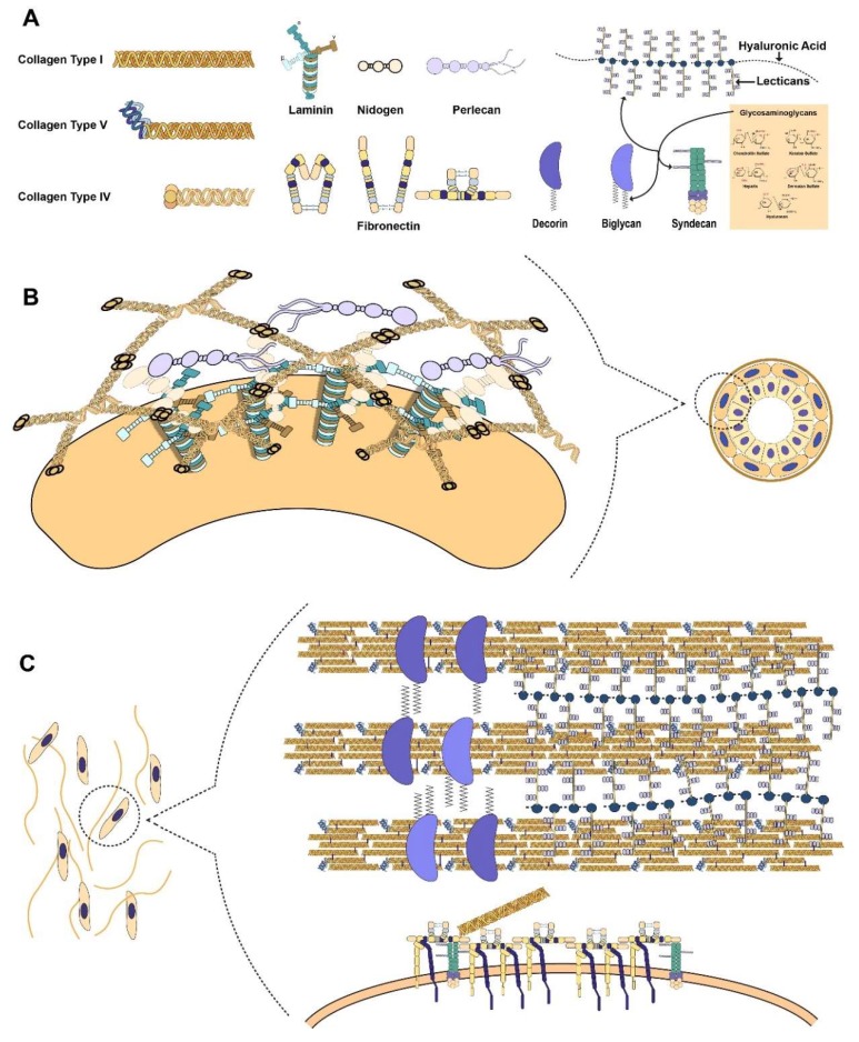
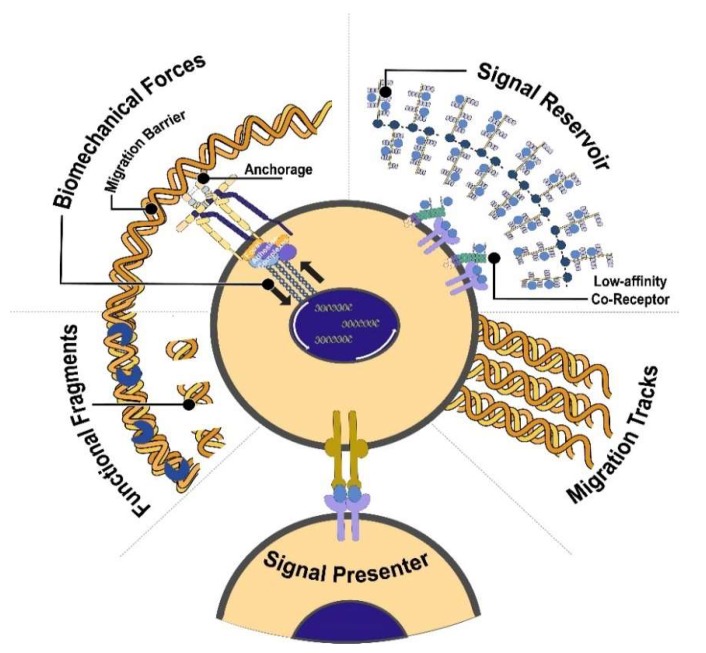
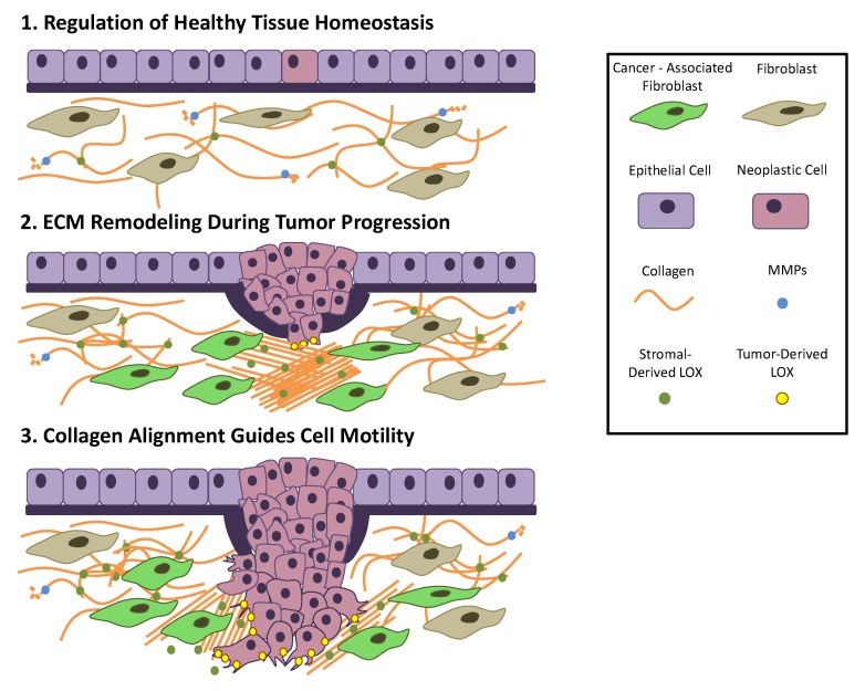
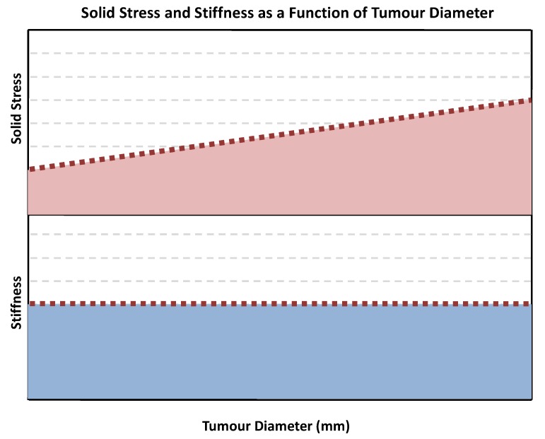
The immense diversity of extracellular matrix (ECM) proteins confers distinct biochemical and biophysical properties that influence cell phenotype. The ECM is highly dynamic as it is constantly deposited, remodelled, and degraded during development until maturity to maintain tissue homeostasis. The ECM's composition and organization are spatiotemporally regulated to control cell behaviour and differentiation, but dysregulation of ECM dynamics leads to the development of diseases such as cancer. The chemical cues presented by the ECM have been appreciated as key drivers for both development and cancer progression. However, the mechanical forces present due to the ECM have been largely ignored but recently recognized to play critical roles in disease progression and malignant cell behaviour. Here, we review the ways in which biophysical forces of the microenvironment influence biochemical regulation and cell phenotype during key stages of human development and cancer progression.

Keywords: cancer progression; extracellular matrix; fibrosis; matrix remodelling; tumour microenvironment.


Conflict of interest statement

The authors declare no conflict of interest.

Figures

Figure 1
Figure 1
Unique ECM molecules and their organization in the basement membrane and interstitial stroma. Panel A (top) shows the unique components of the extracellular matrix. Panel B and C (middle) shows how these different collagens, proteoglycans, laminins, and fibronectin are organized within the basement membrane (B) and interstitial ECM (C). A breast acinus with epithelial cells is surrounded by myoepithelial cells and the basement membrane. In the basement membrane, the laminin is bound to the cell and forms a network through its long arms. It is then connected to the collagen IV network through nidogen and proteoglycans such as perlecan and agrin. Outside the basement membrane is the interstitial ECM where fibroblasts that produce and remodel the ECM can be found. In the interstitial stroma, collagen fibres are made up of fibrils composed of collagen I and collagen V. The different proteoglycans, such as decorin, biglycan, and hyalectans, holds the fibrils together to form a collagen fibre. Fibronectin is bound to the cell via integrins and syndecans. Once fibronectin is unfolded, it reveals cryptic binding sites for heparan sulfate proteoglycans (HSPGs) and collagen. Modified and combined figures from Mouw et al. 2014 [15] and Hohenester and Yurchenco 2013 [34].
Figure 2
Figure 2
Functions of the ECM. The ECM serves as a point of anchorage for the cells that is essential for maintaining tissue polarity and asymmetric stem cell division. Depending on the context, it can impede or facilitate migration. It can sequester growth factors and prevent its free diffusion. Other ECM components can bind growth factors and can serve as co-receptors or signal presenters, which help determine the direction of cell-cell communication. Through the action of metalloproteinases (MMPs), fragments of the ECM can also influence cell behaviour. The physical properties of the ECM can be sensed by focal adhesion complexes, which lead to a variety of changes in cell phenotype such as reorganization of the 3D genome. Figure modified and adopted from Lu, Weaver, and Werb 2012 [90].
Figure 3
Figure 3
ECM remodelling during cancer progression and initiation. (1) Epithelial neoplastic cells proliferate rapidly, inducing strain on the basement membrane. (2) Basement membrane bulges due to mechanical strain. Adjacent cancer-associated fibroblasts increase deposition of collagen. Stromal-derived lysyl oxidase (LOX) aligns collagen. (3) Neoplastic cells breach membrane and migrate along aligned collagen. (Adapted from Lu et al. [6].)
Figure 4
Figure 4
Solid stress and stiffness as a function of tumour diameter (adapted from Nia et al.) [201]. As rigidity of the ECM remains constant, an increase in tumour diameter is associated with increased solid stress within the tumour.
Figure 5
Figure 5
Schematic of oil micro droplet in vivo stress quantification described by Campàs et al. [203]. An oil droplet with calibrated surface tension is injected into living embryonic or cancerous tissue. As cells proliferate, they exert force onto the micro droplet and deform it. Deformations in curvature (red) of the oil droplet are used to calculate anisotropic stress within the tissue.
Figure 6
Figure 6
Illustration of pre-metastatic niche formation in the liver. (1) The primary tumour located in the pancreas emits tumour-derived secreted factors (TDSFs) and extracellular vesicles (EVs). (2) TDSFs and EVs migrate through the vasculature and bone marrow to the secondary organ. While in the bone marrow, TDSFs and EVs recruit bone marrow-derived stem cells (BDSCs), such as hematopoietic stem cells, to the secondary organ site. (3) TDSFs and EVs induce immune cell recruitment and ECM remodelling through LOX and cancer-associated fibroblasts at the pre-metastatic site. [220]

Similar articles

Cited by

References

    1. Frantz C., Stewart K.M., Weaver V.M. The extracellular matrix at a glance. J. Cell Sci. 2010;123:4195–4200. doi: 10.1242/jcs.023820. - DOI - PMC - PubMed
    1. Bonnans C., Chou J., Werb Z. Remodelling the extracellular matrix in development and disease. Nat. Rev. Mol. Cell Biol. 2014;15:786–801. doi: 10.1038/nrm3904. - DOI - PMC - PubMed
    1. Kai F., Laklai H., Weaver V.M. Force matters: Biomechanical regulation of cell invasion and migration in disease. Trends Cell Biol. 2016;26:486–497. doi: 10.1016/j.tcb.2016.03.007. - DOI - PMC - PubMed
    1. Kim S.H., Turnbull J., Guimond S. Extracellular matrix and cell signalling: The dynamic cooperation of integrin, proteoglycan and growth factor receptor. J. Endocrinol. 2011;209:139–151. doi: 10.1530/JOE-10-0377. - DOI - PubMed
    1. Jablonska-Trypuc A., Matejczyk M., Rosochacki S. Matrix metalloproteinases (mmps), the main extracellular matrix (ecm) enzymes in collagen degradation, as a target for anticancer drugs. J. Enzyme Inhib. Med. Chem. 2016;31:177–183. doi: 10.3109/14756366.2016.1161620. - DOI - PubMed

LinkOut - more resources