Review
. 2018 Oct 4;7(10):156.
doi: 10.3390/cells7100156.

The Role of Hydrogen Peroxide in Redox-Dependent Signaling: Homeostatic and Pathological Responses in Mammalian Cells

Affiliations
Review

The Role of Hydrogen Peroxide in Redox-Dependent Signaling: Homeostatic and Pathological Responses in Mammalian Cells

Noemi Di Marzo et al. Cells. .

Abstract

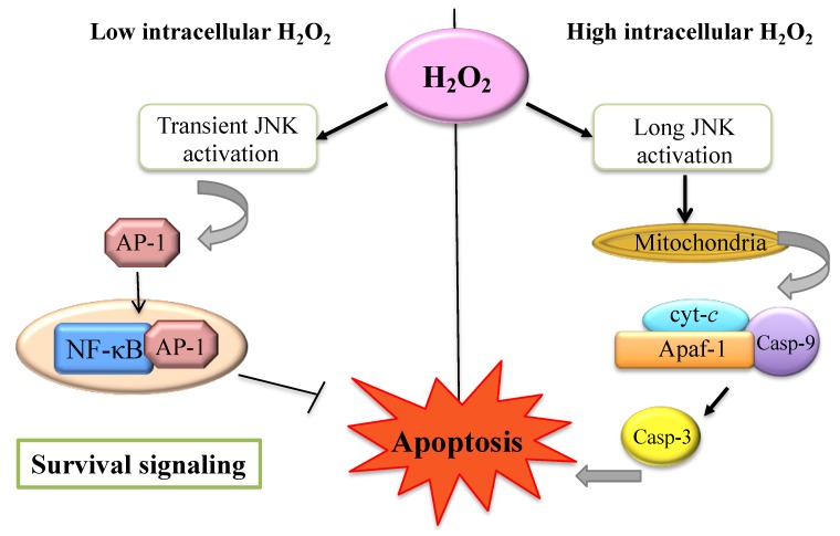
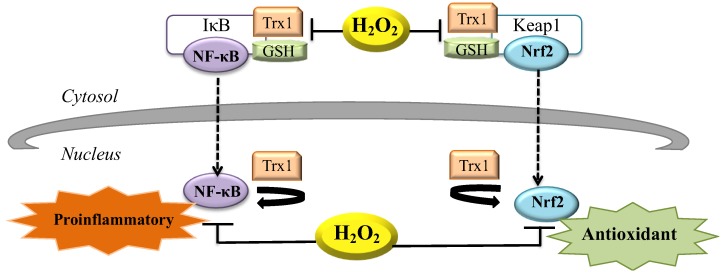
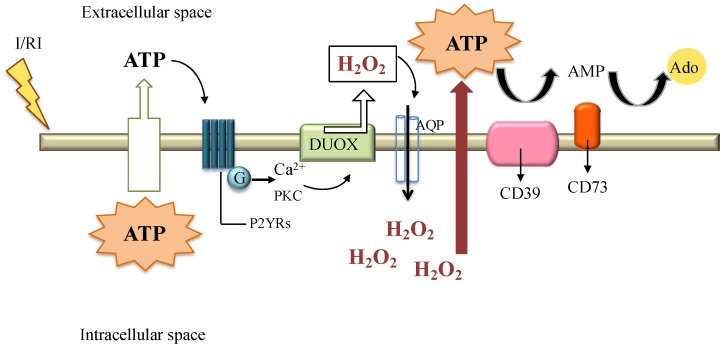
Hydrogen peroxide (H₂O₂) is an important metabolite involved in most of the redox metabolism reactions and processes of the cells. H₂O₂ is recognized as one of the main molecules in the sensing, modulation and signaling of redox metabolism, and it is acting as a second messenger together with hydrogen sulfide (H₂S) and nitric oxide (NO). These second messengers activate in turn a cascade of downstream proteins via specific oxidations leading to a metabolic response of the cell. This metabolic response can determine proliferation, survival or death of the cell depending on which downstream pathways (homeostatic, pathological, or protective) have been activated. The cells have several sources of H₂O₂ and cellular systems strictly control its concentration in different subcellular compartments. This review summarizes research on the role played by H₂O₂ in signaling pathways of eukaryotic cells and how this signaling leads to homeostatic or pathological responses.

Keywords: hydrogen peroxide; oxidative stress; redox regulation.


Conflict of interest statement

The authors declare no conflict of interest.

Figures

Figure 1
Figure 1
H2O2 signaling in mammalian cells. The binding of growth factors (e.g., EGF or PDGF) to their receptors triggers several downstream events. NADPH oxidase (NOX) is a membrane-bound enzyme complex that can produce superoxide anion (O2•−). Activation of this complex (e.g., NOX-2) occurs after the sequential activation of phosphatidylinositol-3-kinase (PI3K) and Rac small GTPase 1 (RAC1) proteins. O2•− produced from NOX complex can dismutate to H2O2 by superoxide dismutase-3 (SOD3). H2O2 can cross the cellular membrane through aquaporin water channels (AQPs) and activates ROS signaling with oxidative modification of critical redox-sensitive Cys in signaling proteins. The targets of H2O2 include transcriptional factors (TFs), mitogen-activated protein kinases (MAPKs) and protein Tyr phosphatases (PTPs). Cellular antioxidant systems, such as catalase (CAT), glutathione peroxidases (GPXs) and peroxiredoxins (PRXs) cooperate to maintain redox homeostasis [9,10].
Figure 2
Figure 2
Survival and apoptotic signaling. High intracellular H2O2 induces long c-Jun NH2-terminal kinase (JNK) activation and lead to mitochondrial cyt-c complex release dependent cell death. Low intracellular H2O2 levels allow AP-1 transcription factor and anti-apoptotic genes activation [40].
Figure 3
Figure 3
Proinflammatory and antioxidant signaling via H2O2. Release of inhibitory subunit (IκB) of NF-κB is due to oxidation by cytosolic H2O2 that leads to dissociation of the NF-κB/IκB complex. Once in the nucleus, NF-κB activity is favored by nuclear thioredoxin-1 (Trx1) and can repair oxidatively damaged proteins. NF-κB activity is inhibited by increased nuclear H2O2 production. During oxidative stress the presence of H2O2 leads to the dissociation of the Nrf2/Keap1 complex and cysteinyl residues of Keap1 are modified. Nrf2 translocates into the nucleus to induce expression of its target genes that triggers antioxidant signaling [59].
Figure 4
Figure 4
Ischemia reperfusion injury (I/RI) induces the passive or active release of intracellular ATP to the extracellular space. Once in the extracellular milieu, ATP can activate G-protein-coupled receptors (P2YRs) to stimulate calcium-dependent signaling and the activation of protein kinase C (PKC) allowing the activation of the DUOX complex and the release of H2O2. H2O2 can cross the cellular membrane via AQP to initiate redox signaling and further promote ATP efflux. Extracellular ATP is metabolized by enzymatic phosphohydrolysis in a two-step process via CD39 conversion of ATP to AMP, and CD73 phosphohydrolysis of AMP to adenosine (Ado). The latter, can mediate anti-inflammatory effects [50,79,84,85].

References

    1. Linley E., Denyer S.P., McDonnell G., Simons C., Maillard J.-Y. Use of hydrogen peroxide as a biocide: New consideration of its mechanisms of biocidal action. J. Antimicrob. Chemother. 2012;67:1589–1596. doi: 10.1093/jac/dks129. - DOI - PubMed
    1. Jones D.P., Sies H. The Redox Code. Antioxid. Redox Signal. 2015;23:734–746. doi: 10.1089/ars.2015.6247. - DOI - PMC - PubMed
    1. Sies H. Hydrogen peroxide as a central redox signaling molecule in physiological oxidative stress: Oxidative eustress. Redox Biol. 2017;11:613–619. doi: 10.1016/j.redox.2016.12.035. - DOI - PMC - PubMed
    1. Lorenzen I., Mullen L., Bekeschus S., Hanschmann E.-M. Redox Regulation of Inflammatory Processes Is Enzymatically Controlled. Oxid. Med. Cell. Longev. 2017;2017:8459402. doi: 10.1155/2017/8459402. - DOI - PMC - PubMed
    1. Veal E.A., Day A.M., Morgan B.A. Hydrogen peroxide sensing and signaling. Mol. Cell. 2007;26:1–14. doi: 10.1016/j.molcel.2007.03.016. - DOI - PubMed

LinkOut - more resources