. 2018 Oct 4;8(1):14798.
doi: 10.1038/s41598-018-33158-8.

Serum GFAP as a biomarker for disease severity in multiple sclerosis

Affiliations

Serum GFAP as a biomarker for disease severity in multiple sclerosis

A Abdelhak et al. Sci Rep. .

Erratum in

Abstract

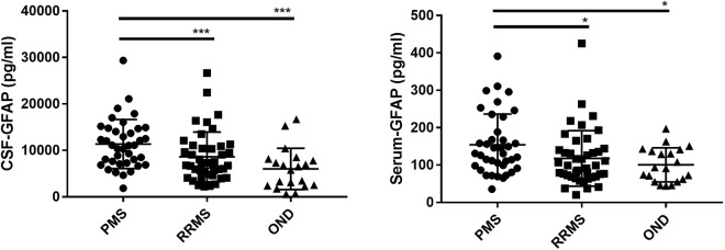
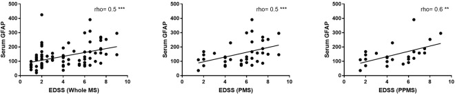
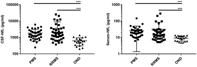
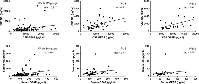
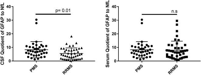
While neurofilament light chain (NfL) measurement in serum is a well-established marker of neuroaxonal damage in multiple sclerosis (MS), data on astroglial markers in serum are missing. In our study, glial fibrillary acid protein (GFAP) and NfL were measured in cerebrospinal fluid (CSF) and serum of MS patients and patients with other non-inflammatory neurological diseases (OND) using the Simoa technology. Clinical data like age, gender, expanded disability status scale (EDSS) and MRI findings were correlated to neurochemical markers. We included 80 MS patients: 42 relapsing-remitting MS (RRMS), 38 progressive MS (PMS), as well as 20 OND. Serum GFAP levels were higher in PMS compared to RRMS and OND (p < 0.001, p = 0.02 respectively). Serum GFAP levels correlated with disease severity in the whole MS group and PMS (Spearman-rho = 0.5, p < 0.001 in both groups). Serum GFAP correlated with serum NfL in PMS patients (Spearman-rho = 0.4, p = 0.01). Levels of serum GFAP were higher with increasing MRI-lesion count (p = 0.01). in summary, we report elevated levels of GFAP in the serum of MS patients. Since serum levels of GFAP correlate with the clinical severity scores and MRI lesion count, especially in PMS patients, it might be a suitable disease progression marker.


Conflict of interest statement

The authors declare no competing interests.

Figures

Figure 1
Figure 1
GFAP CSF and serum levels in multiple sclerosis patients and patients with other non-inflammatory neurological diseases (OND). GFAP: glial fibrillary acidic protein. CSF: cerebrospinal fluid, PMS: progressive multiple sclerosis, RRMS: relapsing-remitting multiple sclerosis. P-values were calculated with Kruskal-Wallis test followed by Dunn’s multiple comparison tests. *p < 0.05; ***p < 0.001.
Figure 2
Figure 2
Spearman correlation (rho) between serum glial fibrillary acidic protein (GFAP) and expanded disability status scale (EDSS) in the whole multiple sclerosis (MS), progressive (PMS) and primary progressive (PPMS) patients. **p < 0.01, ***p < 0.001.
Figure 3
Figure 3
NfL CSF and serum levels in multiple sclerosis patients and patients with other non-inflammatory neurological diseases (OND). NfL: neurofilament light chain, CSF: cerebrospinal fluid, PMS: progressive multiple sclerosis, RRMS: relapsing-remitting multiple sclerosis. OND: other non-inflammatory neurological diseases. P-values were calculated with Kruskal-Wallis test followed by Dunns multiple comparison test. ***p < 0.001.
Figure 4
Figure 4
Spearman correlation (rho) between glial fibrillary acidic protein (GFAP) and neurofilaments light (NfL) in cerebrospinal fluid (CSF) and serum in the whole multiple sclerosis (MS), progressive (PMS) and primary progressive (PPMS) patients. *p < 0.05, **p < 0.01, ***p < 0.001.
Figure 5
Figure 5
Quotient of glial fibrillary acidic protein (GFAP) and neurofilaments light (NfL) in cerebrospinal fluid (CSF) and serum in progressive multiple sclerosis patients (PMS) compared to relapsing-remitting multiple sclerosis (RRMS). n.s.: not significant.

References

    1. Correale J, Farez MF. The Role of Astrocytes in Multiple Sclerosis Progression. Front. Neurol. 2015;6:180. doi: 10.3389/fneur.2015.00180. - DOI - PMC - PubMed
    1. Lassmann H. Multiple sclerosis: lessons from molecular neuropathology. Exp. Neurol. 2014;262(Pt A):2–7. doi: 10.1016/j.expneurol.2013.12.003. - DOI - PubMed
    1. Liddelow SA, et al. Neurotoxic reactive astrocytes are induced by activated microglia. Nature. 2017;541:481–487. doi: 10.1038/nature21029. - DOI - PMC - PubMed
    1. Petzold A, et al. Markers for different glial cell responses in multiple sclerosis: clinical and pathological correlations. Brain: a journal of neurology. 2002;125:1462–1473. doi: 10.1093/brain/awf165. - DOI - PubMed
    1. Axelsson M, et al. Glial fibrillary acidic protein: a potential biomarker for progression in multiple sclerosis. Journal of neurology. 2011;258:882–888. doi: 10.1007/s00415-010-5863-2. - DOI - PubMed