. 2018 Dec;28(12):1171-1185.
doi: 10.1038/s41422-018-0090-y. Epub 2018 Oct 4.

Tom20 senses iron-activated ROS signaling to promote melanoma cell pyroptosis

Affiliations

Tom20 senses iron-activated ROS signaling to promote melanoma cell pyroptosis

Bo Zhou et al. Cell Res. 2018 Dec.

Abstract

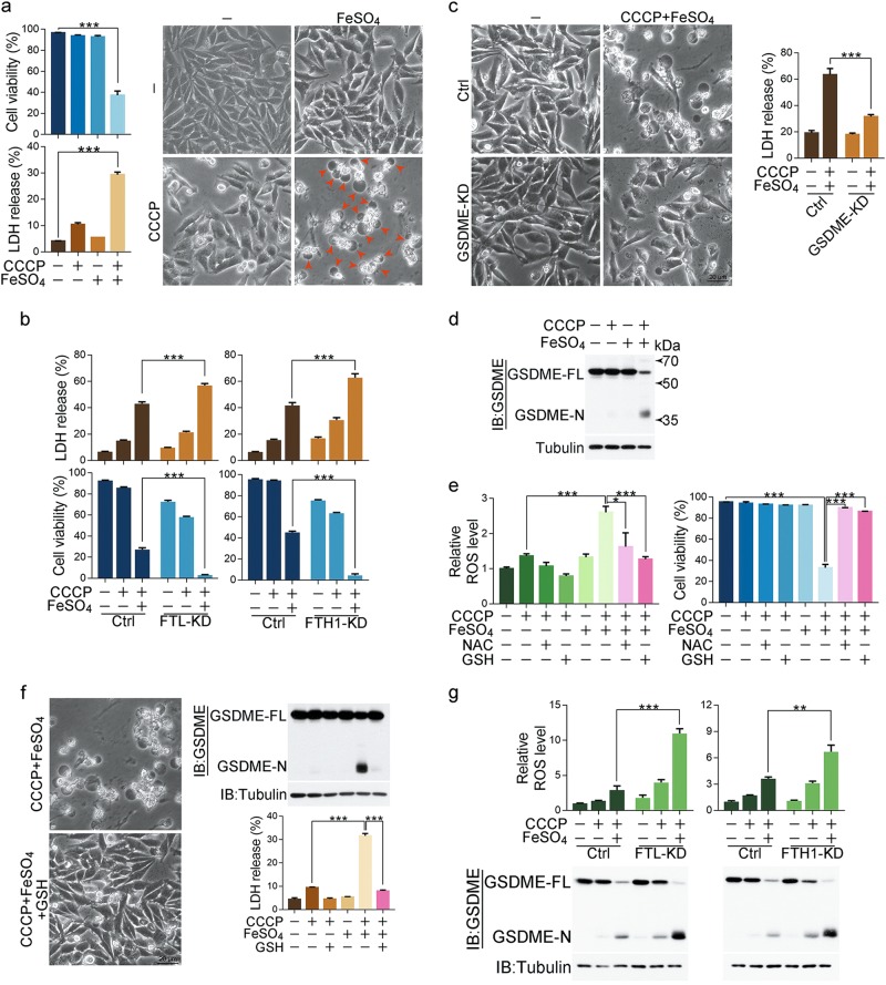
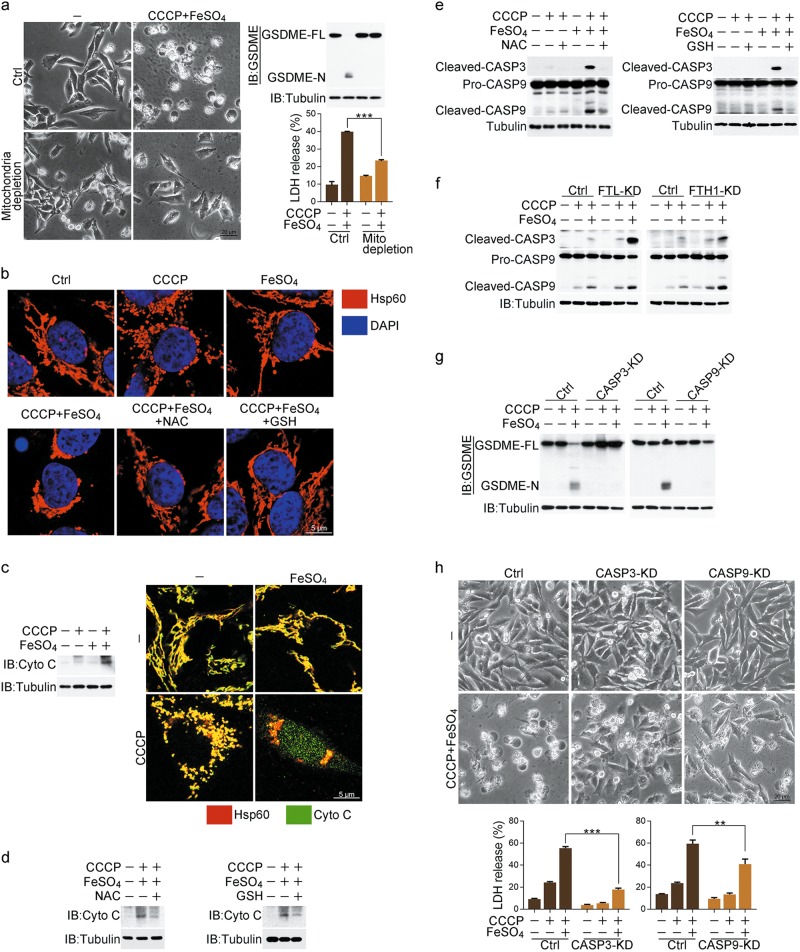
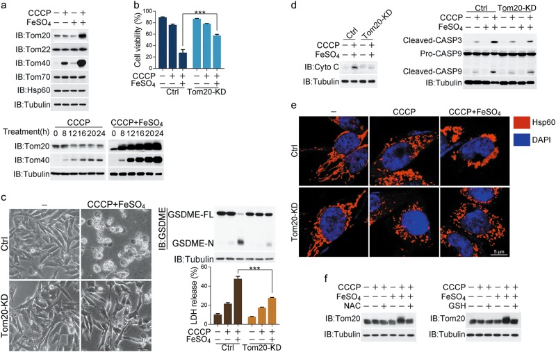
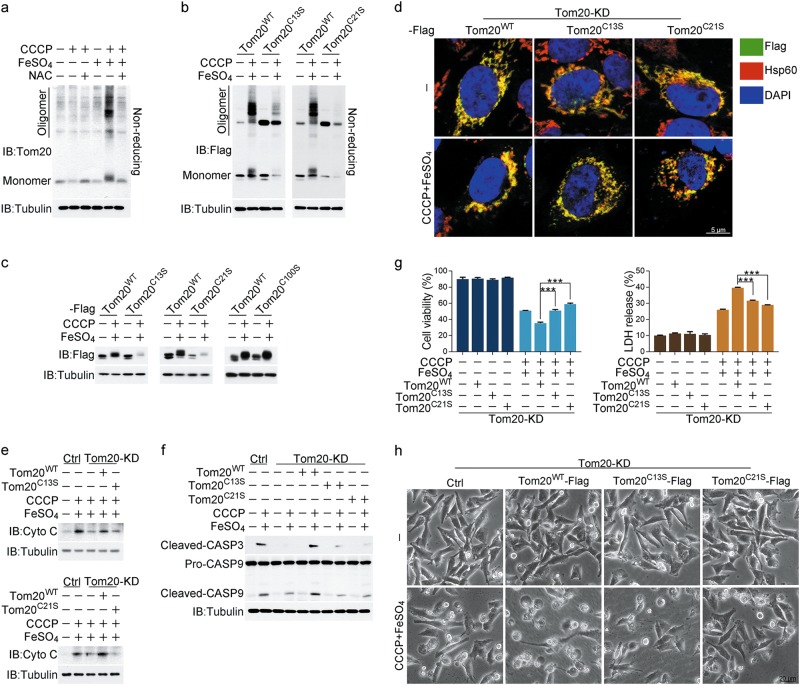
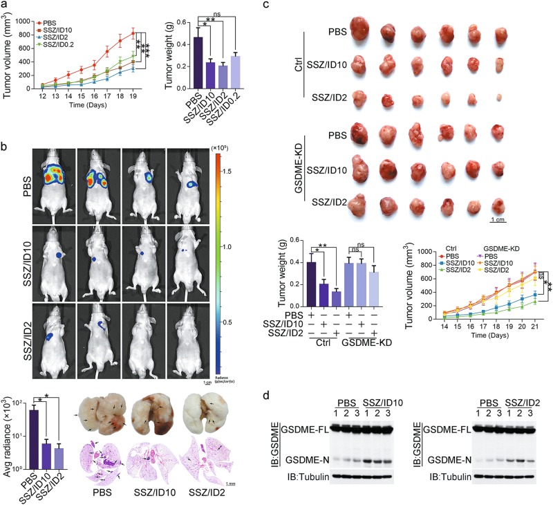
Iron has been shown to trigger oxidative stress by elevating reactive oxygen species (ROS) and to participate in different modes of cell death, such as ferroptosis, apoptosis and necroptosis. However, whether iron-elevated ROS is also linked to pyroptosis has not been reported. Here, we demonstrate that iron-activated ROS can induce pyroptosis via a Tom20-Bax-caspase-GSDME pathway. In melanoma cells, iron enhanced ROS signaling initiated by CCCP, causing the oxidation and oligomerization of the mitochondrial outer membrane protein Tom20. Bax is recruited to mitochondria by oxidized Tom20, which facilitates cytochrome c release to cytosol to activate caspase-3, eventually triggering pyroptotic death by inducing GSDME cleavage. Therefore, ROS acts as a causative factor and Tom20 senses ROS signaling for iron-driven pyroptotic death of melanoma cells. Since iron activates ROS for GSDME-dependent pyroptosis induction and melanoma cells specifically express a high level of GSDME, iron may be a potential candidate for melanoma therapy. Based on the functional mechanism of iron shown above, we further demonstrate that iron supplementation at a dosage used in iron-deficient patients is sufficient to maximize the anti-tumor effect of clinical ROS-inducing drugs to inhibit xenograft tumor growth and metastasis of melanoma cells through GSDME-dependent pyroptosis. Moreover, no obvious side effects are observed in the normal tissues and organs of mice during the combined treatment of clinical drugs and iron. This study not only identifies iron as a sensitizer amplifying ROS signaling to drive pyroptosis, but also implicates a novel iron-based intervention strategy for melanoma therapy.


Conflict of interest statement

The authors declare no competing interests.

Figures

Fig. 1
Fig. 1
Iron activates ROS to promote the pyroptotic death of melanoma cells. Melanoma A375 cells were pretreated with or without different inhibitors, including NAC (5 mM) or GSH (1 mM), for 2 h, followed by CCCP (20 μM), FeSO4 (100 μM), or CCCP/FeSO4 treatment for 6 h to detect the ROS level or 24 h to assess the pyroptotic features (including morphology, GSDME cleavage, and LDH release) and cell viability, unless specifically defined. a The addition of FeSO4 to CCCP induced cell death, LDH release, and pyroptosis (pyroptotic cells are indicated by red arrows). b Knockdown of FTL or FTH1 enhanced LDH release and cell death in response to CCCP/FeSO4 stimulation. KD: knockdown. c Knockdown of GSDME diminished FeSO4-induced pyroptosis and LDH release in the presence of CCCP. d Cleavage of GSDME was observed in response to CCCP/FeSO4 stimulation. e The effects of NAC or GSH on CCCP/FeSO4-induced ROS elevation and cell death. f GSH blocked CCCP/FeSO4-induced pyroptosis, GSDME cleavage and LDH release. g Knockdown of FTL or FTH1 enhanced CCCP/FeSO4-induced LDH release and GSDME cleavage. Tubulin was used to determine the amount of loading proteins. All data are presented as the mean ± SEM of three independent experiments. **P< 0.01, ***P < 0.001
Fig. 2
Fig. 2
Mitochondrial pathway with activation of caspase-3 is involved in pyroptosis induced by iron. Melanoma A375 cells were pretreated with or without different inhibitors, including NAC (5 mM) or GSH (1 mM) for 2 h, followed by CCCP (20 μM), FeSO4 (100 μM), or CCCP/FeSO4 treatment for 6 h to detect cytochrome c release and caspase-3 or -9 cleavage or 24 h to assess the pyroptotic features (including morphology, GSDME cleavage, and LDH release), unless specifically defined. a Mitochondrial depletion blocked CCCP/FeSO4-induced pyroptosis, GSDME cleavage and LDH release. Mito: mitochondria. b CCCP/FeSO4 induced mitochondrial accumulation, but NAC and GSH attenuated this accumulation. c CCCP/FeSO4 induced cytochrome c release from mitochondria to cytosol as detected in the cytosol fraction (left) or by confocal microscopy (right). Cyto C: cytochrome c. d, e NAC or GSH abolished CCCP/FeSO4-induced cytochrome c release (d) and cleavage of caspase-3 and -9 (e). CASP: caspase. f Knockdown of FTL or FTH1 enhanced the CCCP/FeSO4-induced cleavage of caspase-3 and -9. g, h Knockdown of either caspase-3 or -9 abolished the CCCP/FeSO4-induced GSDME cleavage (g), pyroptosis and LDH release (h). Tubulin was used to determine the amount of loading proteins. All data are presented as the mean ± SEM of three independent experiments. **P< 0.01, ***P< 0.001
Fig. 3
Fig. 3
Iron-induced Tom20 accumulation promotes pyroptosis. Melanoma A375 cells were pretreated with or without different inhibitors, including NAC (5 mM) or GSH (1 mM) for 2 h, followed by CCCP (20 μM), FeSO4 (100 μM), or CCCP/FeSO4 treatment for 6 h to detect cytochrome c release and caspase-3 or -9 cleavage or 24 h to assess the pyroptotic features (including morphology, GSDME cleavage, and LDH release) and cell death, unless specifically defined. a Top, addition of FeSO4 to CCCP induced Tom20 and Tom40 accumulation. Bottom, the effect of CCCP/FeSO4 on the expression levels of Tom20 and Tom40. Cells were treated with CCCP or CCCP/FeSO4 for the indicated times. b Knockdown of Tom20 rescued the cell viability in response to CCCP/FeSO4 stimulation. ce Knockdown of Tom20 reversed the CCCP/FeSO4-induced cell morphology from pyroptosis to normal state, reduced LDH release and blocked GSDME cleavage (c), abolished cytochrome c release (detected in the cytosol fraction) and cleavage of caspase-3 and -9 (d), and blocked mitochondria aggregation (e). Hsp60 was used as a mitochondrial indicator, and DAPI was used to display the nuclei. f NAC and GSH attenuated the CCCP/FeSO4-induced Tom20 accumulation. Tubulin was used to determine the amount of loading proteins. Hsp60 was used to determine the amount of mitochondrial proteins. All data are presented as the mean ± SEM of three independent experiments. ***P< 0.001
Fig. 4
Fig. 4
ROS induces Tom20 oxidation in response to iron stimulation. Melanoma A375 cells were pretreated with or without different inhibitors, including NAC (5 mM) for 2 h, followed by CCCP (20 μM), FeSO4 (100 μM), or CCCP/FeSO4 treatment for 6 h to detect cytochrome c release and caspase-3 or -9 cleavage or 24 h to assess the pyroptotic features (including morphology, GSDME cleavage, and LDH release) and cell death, unless specifically defined. To detect the effects of the Tom20 point mutants Tom20C13S, Tom20C21S and Tom20C100S, Tom20 was knocked down in A375 cells, and Tom20WT or its point mutants Tom20C13S, Tom20C21S and Tom20C100S were separately transfected into cells. a FeSO4 induced Tom20 oxidation, which was attenuated by NAC even in the presence of CCCP. Western blotting was performed under reducing and non-reducing conditions to determine the Tom20 oxidation status. b Mutation of either Cys13 or Cys21 in Tom20 abolished the CCCP/FeSO4-induced Tom20 oxidation. c CCCP/FeSO4 could not induce Tom20C13S and Tom20C21S accumulation. d Mutation of either Cys13 or Cys21 in Tom20 blocked mitochondrial aggregation as shown by confocal microscopy. Hsp60 represents the mitochondria, and DAPI was used to display the nuclei. e-h Mutation of either Cys13 or Cys21 in Tom20 abrogated the CCCP/FeSO4-induced cytochrome c release as detected in the cytosol fraction (e), cleavage of caspase-3 and -9 (f), LDH release and cell death (g), and reversed the cell morphology from pyroptosis to normal state (h). Tubulin was used to determine the amount of loading proteins. Hsp60 was used to determine the amount of mitochondrial proteins. All data are presented as the mean ± SEM of three independent experiments. ***P< 0.001
Fig. 5
Fig. 5
Tom20-induced translocation of Bax to mitochondria contributes to pyroptosis. Melanoma A375 cells were treated with CCCP (20 μM), FeSO4 (100 μM), or CCCP/FeSO4 for 6 h to detect the translocation of Bax to mitochondria, cytochrome c release and caspase-3 or -9 cleavage or 24 h to assess the pyroptotic features (including morphology, GSDME cleavage, and LDH release) and cell death, unless specifically defined. To detect the effects of the Tom20 point mutants Tom20C13S and Tom20C21S, Tom20 was knocked down in A375 cells, and Tom20WT or its point mutants Tom20C13S and Tom20C21S were separately transfected into cells. a Bax translocated to mitochondria upon CCCP/FeSO4 stimulation as shown by confocal microscopy. b Knockdown of Tom20 blocked the translocation of Bax to mitochondria upon CCCP/FeSO4 treatment. The mitochondrial fraction in cells was prepared. c Knockdown of Bax blocked the CCCP/FeSO4-induced mitochondrial aggregation. d-g After knocking down Bax, the CCCP/FeSO4-induced cytochrome c release detected in the cytosol fraction was diminished (d), the cleavage of caspase-3 and -9 was attenuated (e), the cell viability was rescued (f), the cell morphologies were reversed from pyroptosis to normal state, and LDH release and GSDME cleavage were also abolished (g). h Effects of the mutants Tom20C13S and Tom20C21S on the translocation of Bax to mitochondria in response to CCCP/FeSO4 stimulation. Hsp60 was used to detect the mitochondria, and DAPI was used to display the nuclei by confocal microscopy. Tubulin was used to determine the amount of loading proteins. Hsp60 was used to determine the amount of mitochondrial proteins. All data are presented as the mean ± SEM of three independent experiments. ***P< 0.001
Fig. 6
Fig. 6
Iron acts as a sensitizer for different drugs and induces pyroptosis in melanoma cells. Melanoma A375 cells were treated with SSZ (sulfasalazine, 125 μM) with or without FeSO4 (100 μM) for 6 h to detect the ROS level or 24 h to assess the pyroptotic features (including morphology, GSDME cleavage, and LDH release), unless specifically defined. a FeSO4 acts as a sensitizer for ROS generation, GSDME cleavage, LDH release, and pyroptosis in the presence of SSZ. b, c Extensive treatment with SSZ/FeSO4 at the indicated times induced Tom20 oxidation and accumulation (b) and GSDME cleavage (c). dg Separate knockdown of Tom20, Bax, caspase-3 or GSDME blocked SSZ/FeSO4-induced pyroptosis, GSDME cleavage, and LDH release as indicated. Tubulin was used to determine the amount of loading proteins. All data are presented as the mean ± SEM of three independent experiments. **P< 0.01, ***P< 0.001
Fig. 7
Fig. 7
Physiological role of iron in suppressing melanoma development. a Melanoma A375 cells were injected subcutaneously into the posterior flanks of nude mice (n = 6). After four days, SSZ (50 mg/kg) with or without iron dextran (10 mg/kg, 2 mg/kg and 0.2 mg/kg, respectively) as indicated was intraperitoneally administered to the mice every other day for 2 weeks. The tumor volume and weight were recorded at the indicated times. ID: iron dextran. PBS: phosphate-buffered saline. b Administration of SSZ/ID inhibited tumor metastasis in mice. Luciferase-expressing A375 cells (2 × 106 cells/mouse) were injected into the tail veins of nude mice (n = 7). Mice were administered with SSZ (50 mg/kg)/ID2 (2 mg/kg) or SSZ (50 mg/kg)/ID10 (10 mg/kg) every other day for 50 days, and tumor metastasis was quantified using bioluminescence imaging. Representative images of mice and luciferase signal intensities were shown. The tumors formed in the lungs were indicated by H&E staining. c Effects of GSDME knockdown on tumor growth, tumor volume and weight. GSDME was knocked down in A375 cells and injected subcutaneously into the posterior flanks of nude mice (n = 6). The mice were treated with the drugs as described above. Images of xenograft tumors in nude mice, tumor weights and volumes were shown. d The cleavage of GSDME was detected in xenograft tumors from the tumor samples (ctrl group) of c
Fig. 8
Fig. 8
A novel pathway of Tom20-Bax-caspase-GSDME upon iron stimulation. In melanoma cells, iron-elevated ROS causes the oxidation and oligomerization of Tom20. Oxidized Tom20 induces Bax translocation to mitochondria, which facilitates cytochrome c release to cytosol. Once released, cytochrome c activates caspase-9, which then activates caspase-3. This caspase-3 activation further cleaves GSDME, and eventually triggers cell swelling and LDH release

References

    1. Sabharwal SS, Schumacker PT. Mitochondrial ROS in cancer: initiators, amplifiers or an Achilles’ heel? Nat. Rev. Cancer. 2014;14:709–721. doi: 10.1038/nrc3803. - DOI - PMC - PubMed
    1. Gorrini C, Harris IS, Mak TW. Modulation of oxidative stress as an anticancer strategy. Nat. Rev. Drug Discov. 2013;12:931–947. doi: 10.1038/nrd4002. - DOI - PubMed
    1. Schieber M, Chandel NS. ROS function in redox signaling and oxidative stress. Curr. Biol. 2014;24:R453–R462. doi: 10.1016/j.cub.2014.03.034. - DOI - PMC - PubMed
    1. Dixon SJ, Stockwell BR. The role of iron and reactive oxygen species in cell death. Nat. Chem. Biol. 2014;10:9–17. doi: 10.1038/nchembio.1416. - DOI - PubMed
    1. Kagan VE, et al. Cytochrome c acts as a cardiolipin oxygenase required for release of proapoptotic factors. Nat. Chem. Biol. 2005;1:223–232. doi: 10.1038/nchembio727. - DOI - PubMed

Publication types

MeSH terms