

Dermatofibrosarcoma Protuberans of the Breast-a Rare Entity
- PMID: 30287997
- PMCID: PMC6154368
- DOI: 10.1007/s13193-017-0684-8
Dermatofibrosarcoma Protuberans of the Breast-a Rare Entity
Abstract
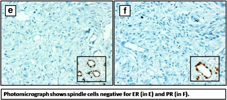
Dermatofibrosarcoma protuberans (DFSP) represents about 1% of soft-tissue sarcomas with an estimated incidence of 0.8 to 5.0 cases per million per year. This lesion may occur anywhere in the body but more than 50% occur on the trunk, 20% on the head and neck and 30% on the extremities. DFSP of the breast is an extremely uncommon site of presentation. Data regarding DFSP of the breast is limited and mostly in the form of case reports. Clinical presentation is not uniform and may mimic benign skin lesions [1]. However, it typically presents as a nodular cutaneous mass in early or mid-adult life. We herein report a case of DFSP of the breast in a 33-year-old lady who was managed successfully in our institute and review the literature associated with it.
Keywords: Breast; Dermatofibrosarcoma protuberans; Lesion.
Figures


References
-
- Nggada HA, Gali BM, Na’aya HU. Clinicopathological study of dermatofibrosarcoma protuberans in Maiduguri, northeastern Nigeria. Niger J Surg Res. 2006;8:78–80.
-
- Weiss SW, Goldblum JR (2008) Extra gastrointestinal stromal tumors. In: Enzinger and Weiss’s Soft Tissue Tumors, 5th edn. Elesevier, Mosby, p 565-79
Publication types
LinkOut - more resources
Full Text Sources