. 2018 Sep 26:10:113-121.
doi: 10.2147/CPAA.S160262. eCollection 2018.

Targeting EGFR and uPAR on human rhabdomyosarcoma, osteosarcoma, and ovarian adenocarcinoma with a bispecific ligand-directed toxin

Affiliations

Targeting EGFR and uPAR on human rhabdomyosarcoma, osteosarcoma, and ovarian adenocarcinoma with a bispecific ligand-directed toxin

Felix Oh et al. Clin Pharmacol. .

Abstract

Purpose: Human sarcomas are rare and difficult to treat cancerous tumors typically arising from soft tissue or bone. Conversely, carcinomas are the most common cancer subtype in humans and the primary cause of mortality across all cancer patients. While conventional therapeutic modalities can prolong disease-free intervals and survival in some cases, treatment of refractory or recurrent solid tumors is challenging, and tumor-related mortality remains unacceptably high. The identification of overexpressed cell surface receptors on sarcoma and carcinoma cells has provided a valuable tool to develop targeted toxins as an alternative anticancer strategy. Recent investigation of recombinant protein-linked toxins that specifically target these cancer receptors has led to the development of highly specific, cytotoxic, and deimmunized drugs that can kill cancer cells.

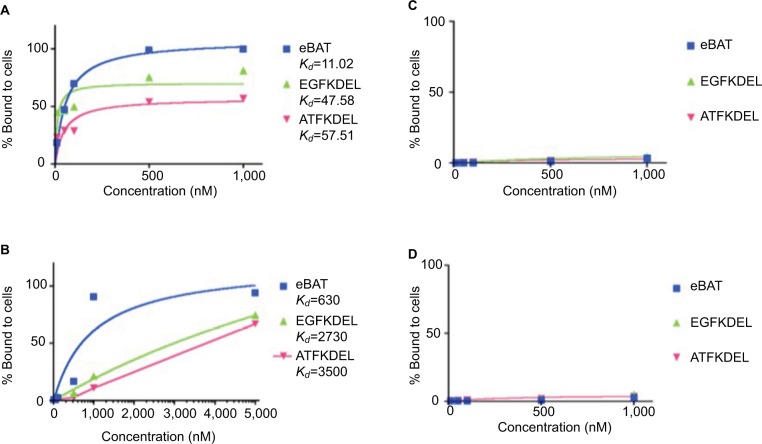
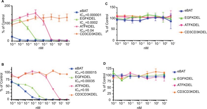
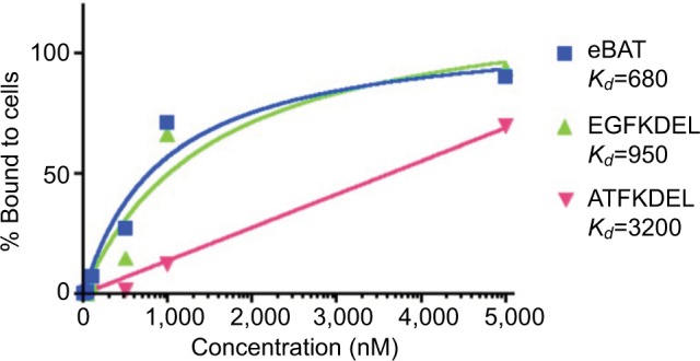
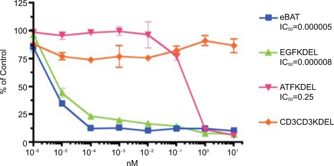
Methods: This study investigated a recombinant protein called epidermal growth factor bispecific angiotoxin (eBAT), which is designed to target the epidermal growth factor receptor (EGFR) on cancer cells and the urokinase plasminogen activator receptor (uPAR) on cancer cells and associated tumor vasculature. Both receptors are expressed by a variety of human sarcomas and carcinomas. Flow cytometry techniques were used to determine binding affinity of eBAT to cancer cells, and proliferation assays were performed to calculate tumor killing ability based on half-maximal inhibitory concentrations.

Results: eBAT demonstrated cytotoxicity against a variety of sarcoma and carcinoma cells that overexpress EGFR and uPAR in vitro and showed greater cell killing ability and binding affinity to cancer cells compared with its monospecific counterparts.

Conclusion: The results of our study are promising, and further studies will be necessary to confirm the applicability of eBAT as a supplementary therapy for a variety of sarcomas, carcinomas, and possibly other refractory malignancies that express EGFR and uPAR.

Keywords: EGFR; carcinoma; eBAT; sarcoma; uPAR.


Conflict of interest statement

Disclosure Dr Vallera is a member of the Oxis Biotech Scientific Advisory Board and holds equity in the company. This relation ship has been reviewed and managed by the University of Minnesota in accordance with its conflict of interest policies. A Borgatti and D Vallera have ownership interest (including patents) in a patent entitled “Reduction of EGFR therapeutic toxicity” filed by the University of Minnesota Office of Technology Commercialization. The other authors report no conflicts of interest in this work.

Figures

Figure 1
Figure 1
Construction and production of eBAT. Notes: In order to construct eBAT, an NcoI/XhoI gene fragment was cloned encoding the genes for the EGF and ATF ligands and the modified KDEL toxin (A). The eBAT gene was inserted into the protein expression vector pET28c and then transfected into E. coli bacteria. The bacteria were then exposed to antibiotics that killed any bacterium that had not undergone transformation. Inclusion bodies, or aggregates of bacterial proteins, were extracted from the bacteria. Our target protein, eBAT, was then isolated, refolded, dialyzed, and purified. The monospecific counterparts EGFKDEL and ATFKDEL and the negative control CD3CD3KDEL were synthesized using similar methods. Gel electrophoresis was used to confirm the size of the reagents (B). Abbreviations: ATF, amino terminal fragment; eBAT, epidermal growth factor bispecific angiotoxin; EGF, epidermal growth factor.
Figure 2
Figure 2
Binding affinity of eBAT to sarcoma (A, B) and B-cell lymphoma (C, D) cell lines. Notes: FACS analysis was performed on the binding of fluorescently labeled eBAT to Saos2 human osteosarcoma cells and RD rhabdomyosarcoma cells. eBAT bound to EGFR- and uPAR-expressing Saos2 cells with very high affinity (>99%) at 500 and 1,000 nM. (A) In comparison, at 1,000 nM EGFKDEL binding peaked at 81.1% while ATFKDEL binding peaked at only 57.2%. eBAT showed greater binding affinity to human osteosarcoma cells than its monospecific counterparts EGFKDEL (Kd=47.58 nM) and ATFKDEL (Kd=57.51 nM) at any given concentration. RD cells exhibited similar binding patterns with the highest binding affinity to eBAT (>90%) at 5,000 nM followed by EGFKDEL and ATFKDEL. (B) The Kd value of eBAT in human rhabdomyosarcoma cells was 630 nM, indicating that it bound more effectively than EGFKDEL (Kd=2,730 nM) and ATFKDEL (Kd=3,500 nM). eBAT was not expected to bind to Raji and Daudi cells because the two cell lines negatively express EGFR and uPAR. eBAT, EGFKDEL, and ATFKDEL did not bind specifically to Raji and Daudi cells at all concentrations tested. (C, D) The graphs show the population of cancer cells relative to control cells as the concentration of each reagent increases. Kd values were not applicable as the reagents did not bind to >50% of the cells at any given concentration and no significant difference in binding was noted between the reagents. Abbreviations: ATF, amino terminal fragment; eBAT, epidermal growth factor bispecific angiotoxin; EGFR, epidermal growth factor receptor; FACS, fluorescence-activated cell sorting; uPAR, urokinase plasminogen activator receptor.
Figure 3
Figure 3
Cytotoxicity of eBAT against sarcoma (A, B) and B-cell lymphoma (C, D) cell lines in vitro. Notes: Proliferation assays were performed with sarcoma cells in the presence of eBAT, EGFKDEL, ATFKDEL, and CD3CD3KDEL as the irrelevant control. The graphs show the population of cancer cells relative to control cells as the concentration of each reagent increases. In Saos2 osteosarcoma cells, eBAT demonstrated significantly increased cytotoxicity (IC50<0.000001) compared with EGFKDEL (P<0.02) and ATFKDEL (P<0.002) even at the lowest concentrations (A). In RD rhabdomyosarcoma cells, eBAT also demonstrated significantly greater cytotoxicity than both EGFKDEL (P<0.001) and ATFKDEL (P<0.0003) (B). Raji and Daudi cells were used as negative control cells due to the lack of EGFR and uPAR expression on the cell surface. (C, D). eBAT was expected to demonstrate minimal cytotoxicity against both Raji and Daudi cells for this reason. eBAT, EGFKDEL, and ATFKDEL had no effect on the proliferation of Raji and Daudi cells and there was no significant difference in cytotoxicity between the reagents. All significance was determined using Student’s t-test and calculated alongside the SD. The error bars indicate standard deviation. Abbreviations: ATF, amino terminal fragment; eBAT, epidermal growth factor bispecific angiotoxin; EGFR, epidermal growth factor receptor; uPAR, urokinase plasminogen activator receptor.
Figure 4
Figure 4
Binding affinity of eBAT to SKOV3 cells. Notes: FACS analysis was performed on the binding of fluorescently labeled eBAT to SKOV3 ovarian adenocarcinoma cells. eBAT bound to EGFR- and uPAR-expressing SKOV3 cells with 93.8% affinity at 5,000 nM. At the same concentration, EGFKDEL binding peaked at 90.2% while ATFKDEL binding peaked at 70.9%. eBAT had a Kd value of 680 nM, showing greater binding affinity to human carcinoma cells than its monospecific counterparts EGFKDEL (Kd=950 nM) and ATFKDEL (Kd=3,200 nM). Abbreviations: ATF, amino terminal fragment; eBAT, epidermal growth factor bispecific angiotoxin; EGFR, epidermal growth factor receptor; FACS, fluorescence-activated cell sorting; uPAR, urokinase plasminogen activator receptor.
Figure 5
Figure 5
Cytotoxicity of eBAT against SKOV3 cells in vitro. Notes: Proliferation assays were performed with SKOV3 ovarian adenocarcinoma cells in the presence of eBAT, EGFKDEL, ATFKDEL, and CD3CD3KDEL as the irrelevant control. The graphs show the population of cancer cells relative to control cells as the concentration of each reagent increases. Despite not being a sarcoma, SKOV3 is demonstrative of the specific cytotoxicity of eBAT against all EGFR- and uPAR-expressing tumors. In SKOV3 cells, eBAT consistently demonstrated equal or greater cytotoxicity than EGFKDEL and significantly greater cytotoxicity than ATFKDEL (P<0.001). The error bars indicate standard deviation. Abbreviations: ATF, amino terminal fragment; eBAT, epidermal growth factor bispecific angiotoxin; EGFR, epidermal growth factor receptor; uPAR, urokinase plasminogen activator receptor.

References

    1. World Health Organization . Classification of tumours. In: Fletcher CDM, Unni KK, Mertens F, editors. Pathology and Genetics of Tumors of Soft Tissue. Lyon, France: IARC Press; 2002. pp. 12–224.
    1. American Cancer Society . Cancer Facts & Figures. Atlanta, Ga: American Cancer Society; 2018. 2018.
    1. Rosenberg SA, Tepper J, Glatstein E, et al. The treatment of soft-tissue sarcomas of the extremities: prospective randomized evaluations of (1) limb-sparing surgery plus radiation therapy compared with amputation and (2) the role of adjuvant chemotherapy. Ann Surg. 1982;196(3):305–315. - PMC - PubMed
    1. Survival rates for ovarian cancer, by stage 2018. [Accessed April 17, 2018]. Available from: https://www.cancer.org/cancer/ovarian-cancer/detection-diagnosis-staging....
    1. Dorsam RT, Gutkind JS. G-protein-coupled receptors and cancer. Nat Rev Cancer. 2007;7(2):79–94. - PubMed