. 2019 Mar;42(3):371-380.
doi: 10.1007/s00270-018-2081-2. Epub 2018 Oct 4.

Uterine Artery Embolization in Women with Symptomatic Cervical Leiomyomata: Efficacy and Safety

Affiliations

Uterine Artery Embolization in Women with Symptomatic Cervical Leiomyomata: Efficacy and Safety

Annefleur M de Bruijn et al. Cardiovasc Intervent Radiol. 2019 Mar.

Abstract

Purpose: To perform an evaluation on safety and efficacy of uterine artery embolization (UAE) in the patients with symptomatic cervical leiomyomata.

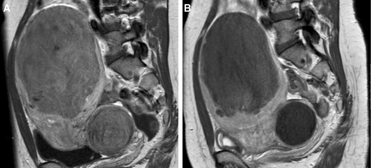
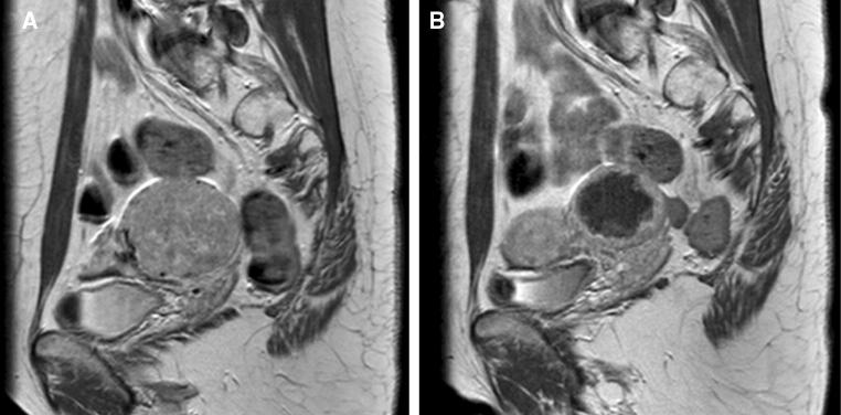
Methods: Patients with symptomatic cervical leiomyomata who underwent UAE in one specialized hospital were retrospectively analyzed, both clinically and with MR imaging. The 3-month outcomes were assessed with MR imaging and a validated questionnaire. Long-term follow-up was assessed by direct contact or file review. To determine the efficacy of UAE for cervical leiomyomata, the primary objective was to assess the clinical outcome with the UFS-QOL questionnaire, containing the health-related quality of life (HRQOL) and symptom severity score (SSS). To assess safety, the secondary objective included leiomyomata volume reduction, the infarction/complication rate and secondary interventions were needed.

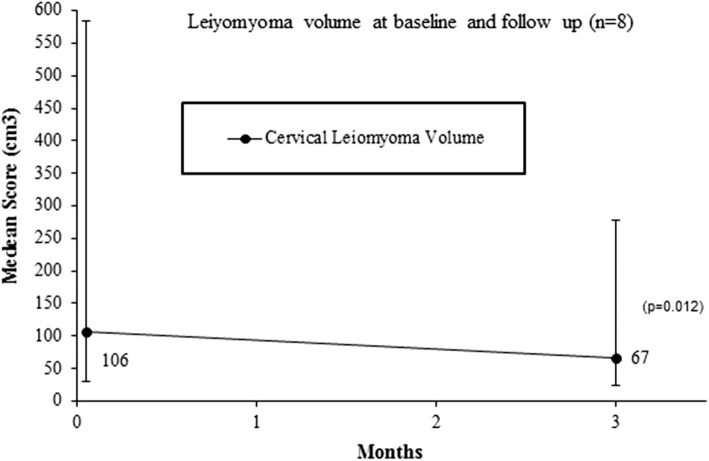
Results: Between 2006 and 2017, eight of 1180 patients underwent UAE and were eligible for inclusion. All embolizations were technically successful (n = 8). At 3 months, all patients showed cervical leiomyomata volume reduction with a median reduction of 41.5% (38.8 cm3) compared to baseline (p = 0.012). No complications occurred. At a median follow-up of 3 months (range 1-7, n = 7), the HRQOL and SSS improved with a median difference of 13 points (range - 5 to 60, p = 0.063) and - 13 points (range - 79 to 3, p = 0.046), respectively. Long-term follow-up showed two secondary interventions (median of 43.5 months). Six patients reported no symptom recurrence.

Conclusion: UAE in women with symptomatic cervical leiomyomata is effective and safe with significant improvement in symptoms and quality of life. UAE is a valuable option for women seeking a non-surgical solution.

Keywords: Cervical leiomyomata; Health-related quality of life (HRQOL); Uterine artery embolization.


Conflict of interest statement

Dr. P.N.M. Lohle has a consultancy agreement with Boston Scientific. All other authors declare that they have no conflicts of interest.

Figures

Fig. 1
Fig. 1
Overview of afferent branching arteries to cervical leiomyomata as identified during UAE. Note Boxes type 1, 2.1 and 2.2 are the locations of the catheters as used per subtype. Subtype 1 with a plexus of feeding branches to the cervical leiomyomata. Subtype 2.1 with a proximal single branch from uterine artery to the cervical leiomyomata. Subtype 2.2 with a distal single branch from uterine artery to the cervical leiomyomata. Type 2 cases were, when possible, super-selectively catheterized
Fig. 2
Fig. 2
Median cervical leiomyomata volume reduction (cm3) until 3 months of follow-up compared to baseline
Fig. 3
Fig. 3
MRI (SAG T1 − TSE − HR contrast +) imaging of cervical leiomyomatas in combination with uterine body fibroids. Note Patient no. 6, MR images (sagittal T1 − TSE − HR + contrast) of leiomyomata in the uterine body and cervix. A Both the cervical leiomyomata (106 cm3) and the uterine body leiomyomata demonstrated full enhancement prior to UAE. B Follow-up imaging 3 months after UAE with 5% enhancement of the leiomyomata in the uterine body and no enhancement of the cervical leiomyomata, as a result of complete infarction after bilateral UAE (2cc700 µm, 4cc900 µm and 4cc1200 µm microspheres, Embozene™). The cervical leiomyomata volume 3 months after UAE was 77 cm3 with a volume reduction of 27%
Fig. 4
Fig. 4
MRI (SAG T1 − TSE − HR contrast +) imaging of a solitary cervical leiomyomata. Note Patient no. 2, MR images (sagittal T1 − TSE − HR + contrast) of a single cervical leiomyomata in a 37-year-old patient with heavy menstrual blood loss and a wish to conceive. Cervix with a broad-based leiomyomata on the left. A The cervical leiomyomata (106 cm3) with full enhancement prior to UAE. B MR imaging 3 months after UAE showed 95% infarction with an irregular enhancing rim. Unilateral left-side UAE with 2cc500, 2cc700 and 3cc900 µm microspheres Embozene™; 47% volume reduction (56.4 cm3)
Fig. 5
Fig. 5
Total quality of life and symptoms severity scores at baseline and 3 months of follow-up

References

    1. Townsend DE, Sparkes RS, Baluda MC, McClelland G. Unicellular histogenesis of uterine leiomyomas as determined by electrophoresis by glucose-6-phosphate dehydrogenase. Am J Obstet Gynecol. 1970;107(8):1168–1173. doi: 10.1016/S0002-9378(15)30365-3. - DOI - PubMed
    1. Buttram VC, Jr, Reiter RC. Uterine leiomyomata: etiology, symptomatology, and management. Fertil Steril. 1981;36(4):433–445. doi: 10.1016/S0015-0282(16)45789-4. - DOI - PubMed
    1. Cramer SF, Patel A. The nonrandom regional distribution of uterine leiomyomas: a clue to histogenesis? Hum Pathol. 1992;23(6):635–638. doi: 10.1016/0046-8177(92)90318-W. - DOI - PubMed
    1. Tiltman AJ. Leiomyomas of the uterine cervix: a study of frequency. Int J Gynecol Pathol. 1998;17(3):231–234. doi: 10.1097/00004347-199807000-00006. - DOI - PubMed
    1. Stovall T. Myomectomy. In: Mann WJ, Stovall T, editors. Gynecologic surgery. New York: Churchill Livingstone; 1996. pp. 445–461.