. 2019 Feb;42(1):185-198.
doi: 10.1007/s10753-018-0883-7.

Alveolar Macrophage Chemokine Secretion Mediates Neutrophilic Lung Injury in Nox2-Deficient Mice

Affiliations

Alveolar Macrophage Chemokine Secretion Mediates Neutrophilic Lung Injury in Nox2-Deficient Mice

Renee M Potera et al. Inflammation. 2019 Feb.

Abstract

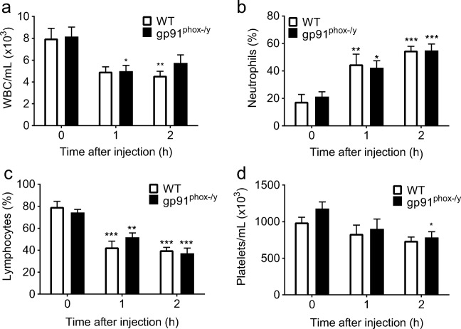
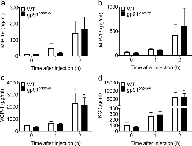
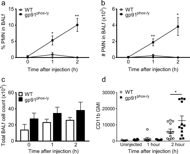
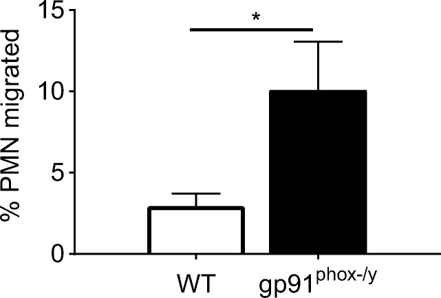
Acute lung injury (ALI), developing as a component of the systemic inflammatory response syndrome (SIRS), leads to significant morbidity and mortality. Reactive oxygen species (ROS), produced in part by the neutrophil NADPH oxidase 2 (Nox2), have been implicated in the pathogenesis of ALI. Previous studies in our laboratory demonstrated the development of pulmonary inflammation in Nox2-deficient (gp91phox-/y) mice that was absent in WT mice in a murine model of SIRS. Given this finding, we hypothesized that Nox2 in a resident cell in the lung, specifically the alveolar macrophage, has an essential anti-inflammatory role. Using a murine model of SIRS, we examined whole-lung digests and bronchoalveolar lavage fluid (BALf) from WT and gp91phox-/y mice. Both genotypes demonstrated neutrophil sequestration in the lung during SIRS, but neutrophil migration into the alveolar space was only present in the gp91phox-/y mice. Macrophage inflammatory protein (MIP)-1α gene expression and protein secretion were higher in whole-lung digest from uninjected gp91phox-/y mice compared to the WT mice. Gene expression of MIP-1α, MCP-1, and MIP-2 was upregulated in alveolar macrophages obtained from gp91phox-/y mice at baseline compared with WT mice. Further, ex vivo analysis of alveolar macrophages, but not bone marrow-derived macrophages or peritoneal macrophages, demonstrated higher gene expression of MIP-1α and MIP-2. Moreover, isolated lung polymorphonuclear neutrophils migrate to BALf obtained from gp91phox-/y mice, further providing evidence of a cell-specific anti-inflammatory role for Nox2 in alveolar macrophages. We speculate that Nox2 represses the development of inflammatory lung injury by modulating chemokine expression by the alveolar macrophage.

Keywords: ARDS; NADPH oxidase 2; Nox2; acute lung injury; alveolar macrophage.


Figures

Fig. 1
Fig. 1
Both genotypes exhibit alterations in circulating white blood cells following induction of sterile inflammation. WT and gp91phox-/y mice demonstrate a reduction in circulating white blood cells and increase in the percentage of circulating neutrophils within 1–2 h of intraperitoneal injection of zymosan (a, b). There is a decrease in the percentage of circulating lymphocytes in both genotypes within 1–2 h after injection (c), with only the gp91phox-/y mice developing decreased platelet counts at 2 h (d). Time 0 represents uninjected control mice. (N = 6–17 per time point from seven independent experiments). *p ≤ 0.05, **p ≤ 0.01, ***p ≤ 0.001 as compared to uninjected mice.
Fig. 2
Fig. 2
Plasma levels of inflammatory cytokines and chemokines do not differ between genotypes. There are no significant differences in plasma levels of MIP-1α (a) or MIP-1β (b) following zymosan injection in either genotype. MCP-1 is increased in both genotypes 2 h after zymosan injection, with no significant difference between genotypes (c). Plasma levels of KC are increased in the gp91phox-/y mice 2 h after zymosan injection, with no significant differences between genotypes (d). Time 0 represents uninjected control mice. (N = 4–5 from four independent experiments). *p ≤ 0.05 as compared to uninjected mice.
Fig. 3
Fig. 3
gp91phox-/y mice develop microthrombi on the surface of the lung. Two hours following the induction of sterile inflammation, there is development of microthrombi on the surface of the lung in gp91phox-/y mice (b) that is not present in WT mice (a).
Fig. 4
Fig. 4
gp91phox-/y mice display increased neutrophil sequestration and activation in the lung. Representative dot plots demonstrate the percentage of lung PMNs following i.p. zymosan injection in WT and gp91phox-/y mice (a). Both WT and gp91phox-/y mice display sequestration of neutrophils within the lung 1 h after intraperitoneal injection of zymosan with further increase by 2 h when compared with uninjected control mice (b, c). There is no significant difference in the total cell count in lung homogenates between genotypes (d). Surface expression of CD11b is upregulated on neutrophils from the whole-lung digest of gp91phox-/y mice 2 h after injection when compared to the WT mice (e). (N = 7–18 from four independent experiments). *p ≤ 0.05.
Fig. 5
Fig. 5
gp91phox-/y mice have enhanced gene expression and protein secretion of inflammatory cytokines and chemokines in whole-lung digests. There is higher gene expression of MIP-1α in unstimulated gp91phox-/y mice compared to WT mice (a), with no significant differences in the expression of MCP-1 (b), MIP-1β (c), or MIP-2 (d). (N = 4–11 from four independent experiments). Levels of MIP-1α and MCP-1 in the supernatant of the whole-lung digest were determined using ELISA in uninjected mice and 1 h and 2 h post injection. The gp91phox-/y mice have increased basal secretion of MIP-1α in the lung as compared to WT mice (e), with no significant differences in protein secretion of MCP-1 (f). (N = 3–10 from five independent experiments). *p ≤ 0.05, **p ≤ 0.01, ***p ≤ 0.001.
Fig. 6
Fig. 6
gp91phox-/y mice display neutrophil recruitment to the alveolar space. The gp91phox-/y mice display increased neutrophil infiltration into the alveolar space as measured in the BALf within 1 h and 2 h of zymosan injection when compared to the WT mice (a, b), with no significant difference in total BAL cell count (c). There is higher cell surface expression of CD11b on neutrophils from the BAL 2 h after injection when compared to WT mice (d) (N = 4–10 from five independent experiments). *p ≤ 0.05, **p ≤ 0.01.
Fig. 7
Fig. 7
gp91phox-/y mice display enhanced basal gene expression of inflammatory chemokines in cells isolated from the BALf. There is enhanced gene expression of MIP-1α, MCP-1, and MIP-2 in uninjected gp91phox-/y mice compared to WT mice, with a trend towards increased MIP-1β gene expression (a). (N = 3–15 from four independent experiments). In the BALf, the gp91phox-/y mice display increased protein secretion of MIP-1α at baseline and 1–2 h following intraperitoneal injection compared to WT mice (b), with no difference in the secretion of MCP-1 (c). (N = 6–17 from 10 to 11 independent experiments). *p ≤ 0.05, **p ≤ 0.01, ***p ≤ 0.001.
Fig. 8
Fig. 8
Alveolar macrophages isolated from gp91phox-/y mice display enhanced gene expression of inflammatory chemokines. There is enhanced gene expression of MIP-1α (a), MIP-2 (b), and MIP-1β (c) in alveolar macrophages isolated from gp91phox-/y mice when compared to WT mice. Peritoneal macrophages isolated from gp91phox-/y mice have enhanced gene expression of MIP-1β compared to WT mice. There is increased protein secretion of MIP-1α in alveolar macrophages isolated from gp91phox-/y mice, with no difference in peritoneal macrophages (d). (N = 4–11 from a minimum of four independent experiments). *p ≤ 0.05, **p ≤ 0.01, ***p ≤ 0.001.
Fig. 9
Fig. 9
Isolated lung PMNs migrate to the gp91phox-/y BALf. There is increased neutrophil migration to the gp91phox-/y BALf compared to WT BALf. (N = 5–6 from three independent experiments). *p ≤ 0.05.

References

    1. Matthay MA, Kallet RH. Prognostic value of pulmonary dead space in patients with the acute respiratory distress syndrome. Critical Care. 2011;15(5):185. doi: 10.1186/cc10346. - DOI - PMC - PubMed
    1. Rubenfeld GD, Caldwell E, Peabody E, Weaver J, Martin DP, Neff M, Stern EJ, Hudson LD. Incidence and outcomes of acute lung injury. The New England Journal of Medicine. 2005;353(16):1685–1693. doi: 10.1056/NEJMoa050333. - DOI - PubMed
    1. Bunnell E, Pacht ER. Oxidized glutathione is increased in the alveolar fluid of patients with the adult respiratory distress syndrome. The American Review of Respiratory Disease. 1993;148(5):1174–1178. doi: 10.1164/ajrccm/148.5.1174. - DOI - PubMed
    1. Chow CW, Herrera Abreu MT, Suzuki T, Downey GP. Oxidative stress and acute lung injury. American Journal of Respiratory Cell and Molecular Biology. 2003;29(4):427–431. doi: 10.1165/rcmb.F278. - DOI - PubMed
    1. Matthay MA, Geiser T, Matalon S, Ischiropoulos H. Oxidant-mediated lung injury in the acute respiratory distress syndrome. Critical Care Medicine. 1999;27(9):2028–2030. doi: 10.1097/00003246-199909000-00055. - DOI - PubMed

LinkOut - more resources