. 2018 Dec;176(12):2587-2594.
doi: 10.1002/ajmg.a.40526. Epub 2018 Oct 5.

Preliminary observations of mitochondrial dysfunction in Prader-Willi syndrome

Affiliations

Preliminary observations of mitochondrial dysfunction in Prader-Willi syndrome

Merlin G Butler et al. Am J Med Genet A. 2018 Dec.

Abstract

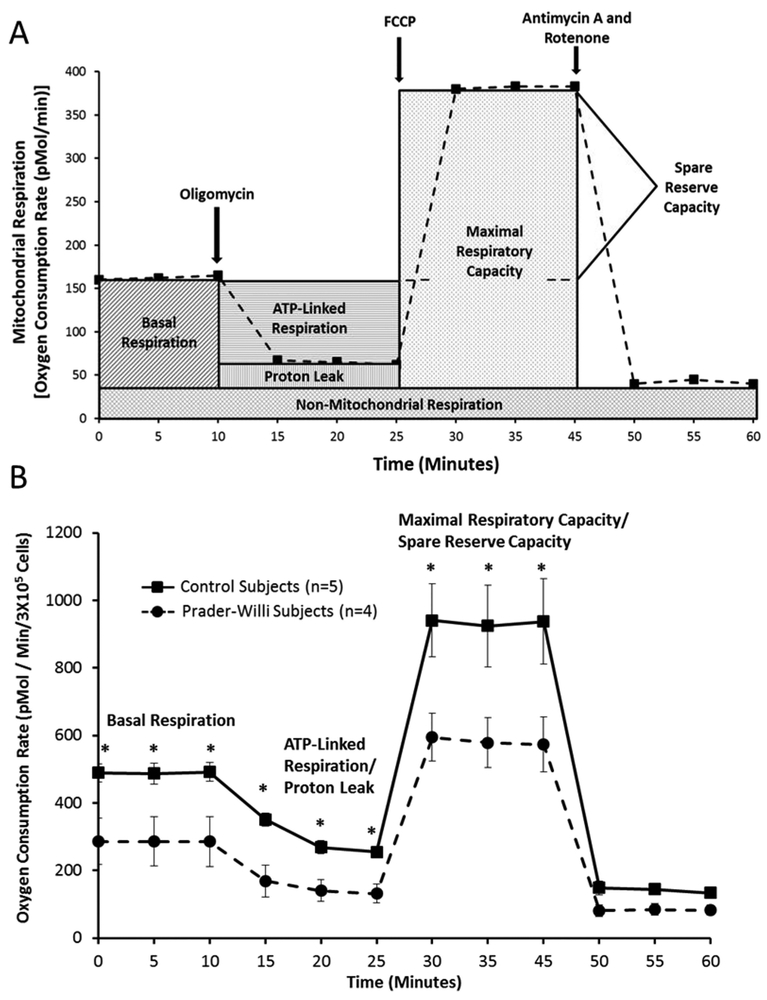
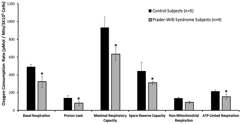
Prader-Willi syndrome (PWS) is a complex multisystem disorder because of errors in genomic imprinting with severe hypotonia, decreased muscle mass, poor suckling, feeding problems and failure to thrive during infancy, growth and other hormone deficiency, childhood-onset hyperphagia, and subsequent obesity. Decreased energy expenditure in PWS is thought to contribute to reduced muscle mass and physical activity but may also relate to cellular metabolism and disturbances in mitochondrial function. We established fibroblast cell lines from six children and adults with PWS and six healthy controls for mitochondrial assays. We used Agilent Seahorse XF extracellular flux technology to determine real-time measurements of several metabolic parameters including cellular substrate utilization, Adenosine Triphosphate (ATP)-linked respiration, and mitochondrial capacity in living cells. Decreased mitochondrial function was observed in the PWS patients compared to the healthy controls with significant differences in basal respiration, maximal respiratory capacity, and ATP-linked respiration. These results suggest disturbed mitochondrial bioenergetics in PWS although the low number of studied subjects will require a larger subject population before a general consensus can be reached to identify if mitochondrial dysfunction is a contributing factor in PWS.

Keywords: Prader-Willi syndrome; fibroblasts; healthy controls; mitochondrial assays and dysfunction.


Conflict of interest statement

There are no conflicts of interest to report for any of the authors.

Figures

Figure 1.
Figure 1.
1A Schematic diagram of Agilent Seahorse XF extra-cellular flux technology and mitochondrial function components and profiles modified from Agilent website (www.agilent.com/en/products/cell-analysis-(seahorse)/mitochondrial-respiration-xf-cell-mito-stress-test). 1B Comparison of stages of mitochondrial function and cellular respiration with time using fibroblasts from Prader-Willi syndrome and control subjects with error bars and significant differences (t test; p<0.05) noted (*) for basal respiration, ATP-linked respiration/proton leak and maximal respiratory capacity with decreased values for Prader-Willi syndrome.
Figure 2.
Figure 2.
Comparison of mitochondrial function and cellular respiration in fibroblasts from Prader-Willi syndrome and control subjects with error bars showing individual parameters for basal respiration, proton leak, maximal respiratory capacity, spare reserve capacity, non-mitochondrial respiration and ATP-linked respiration or coupled respiration. Significant differences (t test; p<0.05) are noted (*) with decreased values in subjects with Prader- Willi syndrome for five of six parameters.
Figure 3.
Figure 3.
Comparison of Prader-Willi syndrome genetic subtypes with p values generated using t tests indicated above the vertical bars.
Figure 4.
Figure 4.
Comparison of mitochondrial function and cellular respiration in fibroblasts from Prader-Willi syndrome (PWS) and control subjects with error bars showing individual parameters for basal respiration, proton leak, maximal respiratory capacity, spare reserve capacity, non-mitochondrial respiration and ATP-linked respiration or coupled respiration. 4A Comparison of control and PWS subjects; 4B Comparison of age and gender matched adult subjects and 4C Comparison of age and gender matched children. Significant differences (t test; p<0.05) are noted (*) with decreased mitochondrial measures in subjects with Prader-Willi syndrome.

Similar articles

Cited by

References

    1. Ainscow EK, & Brand MD (1995). Top‐down control analysis of systems with more than one common intermediate. European Journal of Biochemistry, 231(3), 579–586. - PubMed
    1. Alsaif M, Elliot SA, MacKenzie ML, Prado CM, Field CJ, & Haqq AM (2017). Energy Metabolism Profile in Individuals with Prader-Willi Syndrome and Implications for Clinical Management: A Systematic Review. Advances in Nutrition, 8(6), 905–915. - PMC - PubMed
    1. Angulo M, Butler MG, & Cataletto M (2015). Prader-Willi syndrome: a review of clinical, genetic, and endocrine findings. Journal of Endocrinological Investigation, 38(12), 1249–1263. - PMC - PubMed
    1. Ashley N, O’rourke A, Smith C, Adams S, Gowda V, Zeviani M, . . . Poulton J (2008). Depletion of mitochondrial DNA in fibroblast cultures from patients with POLG1 mutations is a consequence of catalytic mutations. Human Molecular Genetics, 17(16), 2496–2506. - PMC - PubMed
    1. Bittel DC, Kibiryeva N, Talebizadeh Z, & Butler MG (2003). Microarray analysis of gene/transcript expression in Prader-Willi syndrome: Deletion versus UPD. Journal of Medical Genetics, 40(8), 568–574. - PMC - PubMed

Publication types

Substances