Review
. 2018 Oct 4;175(2):327-345.
doi: 10.1016/j.cell.2018.09.021.

The Cellular and Molecular Basis for Planarian Regeneration

Affiliations
Review

The Cellular and Molecular Basis for Planarian Regeneration

Peter W Reddien. Cell. .

Abstract

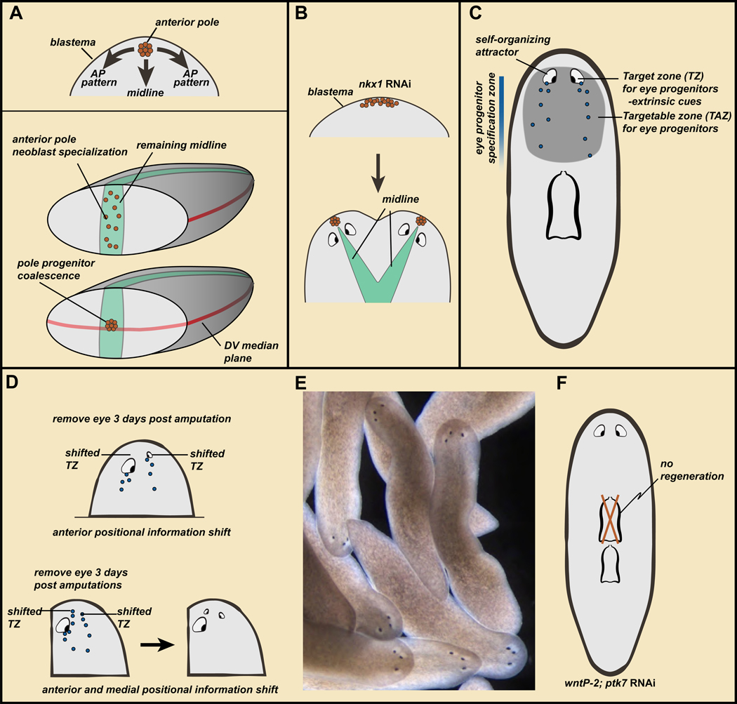
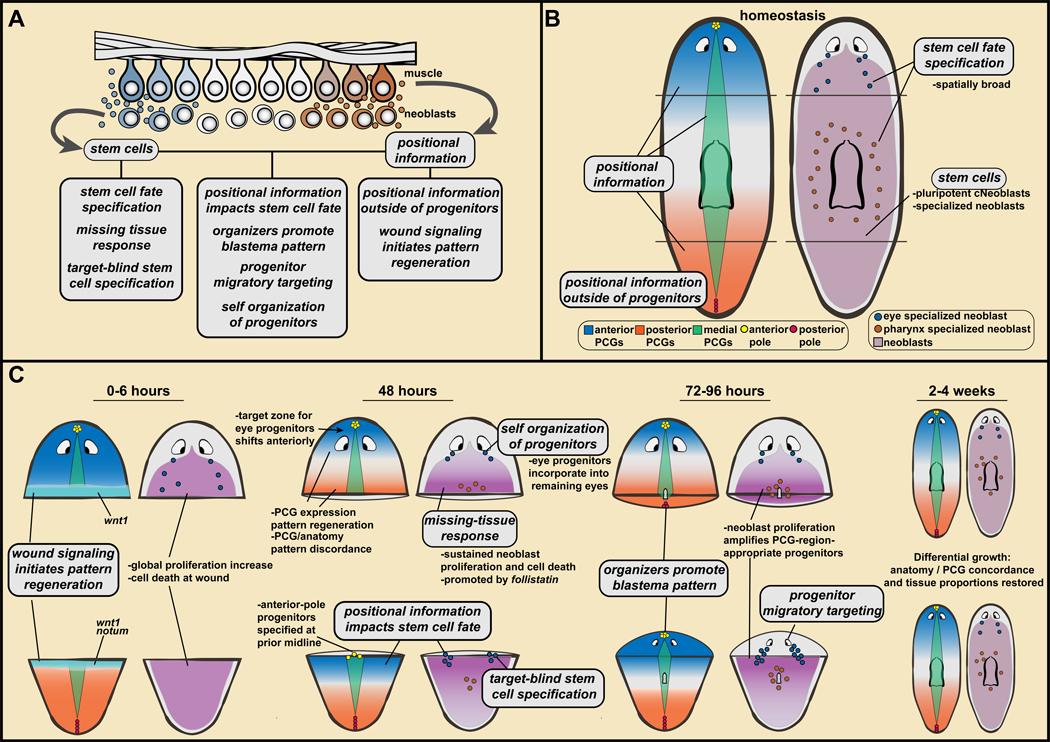
Regeneration is one of the great mysteries of biology. Planarians are flatworms capable of dramatic feats of regeneration, which have been studied for over 2 centuries. Recent findings identify key cellular and molecular principles underlying these feats. A stem cell population (neoblasts) generates new cells and is comprised of pluripotent stem cells (cNeoblasts) and fate-specified cells (specialized neoblasts). Positional information is constitutively active and harbored primarily in muscle, where it acts to guide stem cell-mediated tissue turnover and regeneration. I describe here a model in which positional information and stem cells combine to enable regeneration.


Figures

Figure 1.
Figure 1.. Planarian regeneration.
A–E. Planarian fragments regenerate missing body parts in outgrowths at the wound called blastemas (lighter pigmentation) and through changes in pre-existing tissues (morphallaxis). B. Regeneration from a fragment results in a small animal that can eat and grow towards the original size. C. Parasagittal amputation is followed with ML axis regeneration. D. Incisions not allowed to seal result in duplications of body regions. E. Intercalary regeneration between juxtaposed body fragments.
Figure 2.
Figure 2.. Neoblasts provide the cellular basis for new tissue production in planarian regeneration.
A. Neoblasts (red) are mesenchymal cells distributed broadly. Blue, DAPI; the brain and pharynx are readily visible. From (Wagner et al., 2012). B. Transplantation of single neoblasts can generate colonies with broad differentiation potential and capacity to restore regeneration to lethally irradiated hosts. Resultant animals are genetic clones of the donor (top, right). Bottom right, clonogenic neoblasts (cNeoblasts) are pluripotent stem cells that provide the cellular basis for planarian regeneration. C. Top, specialized neoblasts for the eye produce progenitors that migrate in two trails from the wound into the head blastema, where they coalesce into eyes. Bottom, many specialized neoblast classes produce the differentiated cells of the blastema. The fate of blastema cells is pre-determined based upon which specialized neoblasts they came from. D. Specialized neoblast model. Activation of distinct transcription factors in neoblasts specifies their fate in tissue turnover and regeneration. E. Left: Eyes can be regenerated following resection, but this does not trigger amplification of eye progenitors (magenta; data from (LoCascio et al., 2017)). Right, “target-blind” regeneration: regeneration occurs as an emergent property of constant progenitor production. During regeneration, with no progenitor production-rate change, there are fewer cells in the forming eye accessible to undergo cell death and they are young. Consequently, fewer cell death events per eye passively enable regeneration.
Figure 3.
Figure 3.. Position-control genes are constitutively and regionally expressed in planarian muscle to control adult body plan maintenance and regeneration.
A. Regeneration-patterning phenotypes. Left, trunk fragments regenerating heads and tails. Inhibition of Wnt pathway genes results in regeneration of posterior-facing heads. Inhibition of negative Wnt pathway regulators leads to regeneration of anterior-facing tails. Right, Bmp pathway inhibition leads to dorsal appearance of ventral attributes. B. PCG expression domains. Anterior, up. Data from (Scimone et al., 2016). C. Cartoon map of PCG transcription domains on the AP axis. Blue, Wnt-signaling related; Orange, FGFRL genes; Red, Hox genes; Purple, anterior transcription factors; Grey, unknown molecular function. Bottom: posterior-to-anterior gradient of β-catenin-1 protein level. D. Cartoon of PCG expression domains on the DV/ML axes. Bmp signaling is active dorsally and wnt5 - slit mutual inhibition mediates ML pattern.
Figure 4.
Figure 4.. Wound signaling mediates the regeneration of positional information to enable regeneration of missing parts.
A. PCGs are expressed in muscle. Data from (Witchley et al., 2013). Left, a PCG RNA probe pool (red) labels almost all body-wall muscle cells (green, collagen probe). Right, sFRP-2 transcripts around a muscle cell nucleus. Bar, 20 microns. B. Regeneration model: muscle ßPCG expression, specialized neoblasts, and wound-induced re-establishment of PCG expression domains. See text for details. C. notum is preferentially wound-induced at anterior-facing wounds and wnt1 is wound-induced at all wounds. notum inhibits Wnt signaling to promote head regeneration. How notum is selectivity activated at anterior-facing wounds is unknown. D. Without PCG pattern regeneration in follistatin or myoD RNAi animals, regeneration fails to occur. This requires longitudinal muscle fibers (data from (Scimone et al., 2017)). E. MEK inhibitor treatment blocks regeneration. Removal of the inhibitor does not lead to regeneration unless new injuries are inflicted. F. Coincidence of increased neoblast proliferation with positional information leads to amplification of regional progenitor classes, even if the target tissue of those progenitors remains uninjured.
Figure 5.
Figure 5.. Wound architecture, migratory cues extrinsic to progenitors, and self-organization produce blastema pattern.
A. Top, the anterior pole promotes ML and AP head-blastema pattern. Bottom, wound architectural cues determine the point of anterior pole formation, connecting blastema pattern to the pattern of pre-existing tissue. B. nkx1 RNAi can result in wider initial anterior pole formation in the blastema, with subsequent pole splitting and two heads. C. Model: self-organization and extrinsic targeting cues govern the migratory behavior of progenitors in regeneration. See text for details. D. Amputations lead to PCG shifting, but the self-organizing nature of the eye prevents progenitors from reaching their target zone unless the eye is removed (top) or progenitors are targeted enough medially to miss the eye on their migration path (bottom). E. Wild-type animals with three eyes stably maintain ectopic eyes. Data from (Atabay et al., 2018). F. wntP-2; ptk7 RNAi animals develop a second pharynx; the original anterior pharynx is maintained but not regenerated.
Figure 6.
Figure 6.. A model for planarian regeneration involving positional information and stem cells.
A. Pillars of a model for planarian regeneration are described in text section IV. These pillars refer to stem cell and positional information properties, and how these properties together lead to key steps of regeneration. B, C. Application of pillars to explain head and tail regeneration (also, see text).

References

    1. Abnave P, Aboukhatwa E, Kosaka N, Thompson J, Hill MA, and Aboobaker AA (2017). Epithelial-mesenchymal transition transcription factors control pluripotent adult stem cell migration in vivo in planarians. Development 144, 3440–3453. - PMC - PubMed
    1. Adell T, Saló E, Boutros M, and Bartscherer K. (2009). Smed-Evi/Wntless is required for beta-catenin-dependent and -independent processes during planarian regeneration. Development 136, 905–910. - PubMed
    1. Adler CE, and Sánchez Alvarado A. (2015). Types or States? Cellular Dynamics and Regenerative Potential. Trends Cell Biol 25, 687–696. - PMC - PubMed
    1. Adler CE, Seidel CW, McKinney SA, and Sánchez Alvarado A. (2014). Selective amputation of the pharynx identifies a FoxA-dependent regeneration program in planaria. Elife 3, e02238. - PMC - PubMed
    1. Agata K, Tanaka T, Kobayashi C, Kato K, and Saitoh Y. (2003). Intercalary regeneration in planarians. Dev Dyn 226, 308–316. - PubMed

Publication types

LinkOut - more resources