. 2018 Oct 4;103(4):621-630.
doi: 10.1016/j.ajhg.2018.09.001.

Mutations in KCNK4 that Affect Gating Cause a Recognizable Neurodevelopmental Syndrome

Affiliations

Mutations in KCNK4 that Affect Gating Cause a Recognizable Neurodevelopmental Syndrome

Christiane K Bauer et al. Am J Hum Genet. .

Abstract

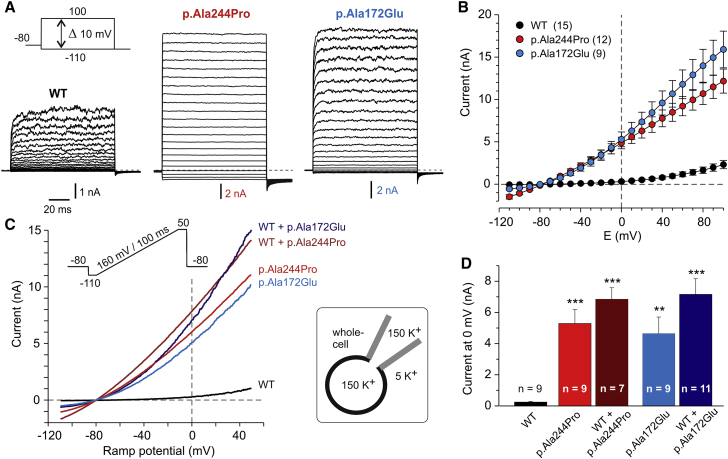
Aberrant activation or inhibition of potassium (K+) currents across the plasma membrane of cells has been causally linked to altered neurotransmission, cardiac arrhythmias, endocrine dysfunction, and (more rarely) perturbed developmental processes. The K+ channel subfamily K member 4 (KCNK4), also known as TRAAK (TWIK-related arachidonic acid-stimulated K+ channel), belongs to the mechano-gated ion channels of the TRAAK/TREK subfamily of two-pore-domain (K2P) K+ channels. While K2P channels are well known to contribute to the resting membrane potential and cellular excitability, their involvement in pathophysiological processes remains largely uncharacterized. We report that de novo missense mutations in KCNK4 cause a recognizable syndrome with a distinctive facial gestalt, for which we propose the acronym FHEIG (facial dysmorphism, hypertrichosis, epilepsy, intellectual disability/developmental delay, and gingival overgrowth). Patch-clamp analyses documented a significant gain of function of the identified KCNK4 channel mutants basally and impaired sensitivity to mechanical stimulation and arachidonic acid. Co-expression experiments indicated a dominant behavior of the disease-causing mutations. Molecular dynamics simulations consistently indicated that mutations favor sealing of the lateral intramembrane fenestration that has been proposed to negatively control K+ flow by allowing lipid access to the central cavity of the channel. Overall, our findings illustrate the pleiotropic effect of dysregulated KCNK4 function and provide support to the hypothesis of a gating mechanism based on the lateral fenestrations of K2P channels.

Keywords: FHEIG syndrome; K2P channels; TRAAK; channelopathy; epilepsy; gingival overgrowth; hypertrichosis; intellectual disability; neurodevelopmental disorder.


Figures

Figure 1
Figure 1
Clinical Features of Individuals with De Novo KCNK4 Mutations (A–C) Subject 1 (11 months). Note the hypotonic face, bitemporal narrowing, bushy and straight eyebrows, long eyelashes, short deep philtrum, prominent upper and lower vermilion, and receding chin. (D–F) Subject 2 at 12 months and 5 years. Evolution of facial appearance is appreciable. Note the bitemporal narrowing, bushy and straight eyebrows, long eyelashes, low-set anteverted ears, short deep philtrum, and prominent upper and lower vermilion. (G–I) Subject 3 at 8 years. Long eyebrows with mild synophris and long eyelashes, thick hair, large mouth with smooth philtrum, and thin lips can be appreciated. Gingival overgrowth and generalized hypertrichosis occur in all affected individuals with different degree of severity.
Figure 2
Figure 2
KCNK4 Structure and Location and Structural Consequences of Disease-Causing Mutations (A) KCNK4 is a homodimeric channel. The schematic structure of the monomer is shown with the cytoplasmic side pointing down (left). Each subunit comprises an extracellular cap covering the pore, four transmembrane helices (M1 to M4), and two pore domains (P1 and P2) constituting the selectivity filter. K+ ions are shown as white spheres. Locations of mutated residues are highlighted by pink spheres. The crystallographic structure of the KCNK4 dimer (PDB: 4WFF) is also shown (lateral view, middle; as seen from the cytoplasm, right). One monomer is colored with the same code of the scheme, while the other monomer is reported in gray. (B) Structure of the KCNK4 channel in the two conformational states with open and closed lateral fenestrations (PDB: 4WFF and 4WFE, left and right panels, respectively). Only helix M4 and residue Ala244 are highlighted in dark red and pink, respectively. K+ ions are shown as white spheres, while the decane molecule inserted in the lateral intramembrane fenestration and blocking the channel is shown as blue spheres. The protein is reported in both ribbon and surface representations, to illustrate the open and closed states of the fenestration that allow or avoid the entrance of the aliphatic molecule. (C) Structural impact of the p.Ala244Pro and p.Ala172Glu amino acid substitutions, as observed in the molecular dynamics (MD) simulations. Histograms of the pore radius of the two lateral openings, observed in the MD simulations (p.Ala244Pro [green], p.Ala172Glu [orange], and WT [red]) (left). The vertical dashed line indicates the radius of a methane molecule (∼2.0 Å). Lateral opening in the conformation that best represents those observed in the MD simulations are also shown (right). The fenestration is viewed from the lipid-exposed external surface and helices M4 and M2 are shown in transparency. Residues Leu125, Phe246, and Leu250 involved in the hydrophobic cluster occluding the fenestration are shown in ball-and-stick representation.
Figure 3
Figure 3
Whole-Cell Current Recordings from CHO Cells Expressing Wild-Type or Mutant KCNK4 Channels (A) Representative current recordings elicited with the indicated pulse protocol consisting of 100 ms voltage steps to potentials between −110 and 100 mV in cells expressing the wild-type (WT) or mutant (p.Ala244Pro or p.Ala172Glu) KCNK4 channels. (B) The I/V-plot shows means ± SEM of current amplitudes recorded with the voltage step protocol. Number of experiments is given in parentheses. Cell transfections were performed using a 1:10 predilution of KCNK4 cDNA (final concentration 400 ng/mL). (C) I/V-plots of representative KCNK4 current recordings elicited with the ramp protocol shown as inset. Cells were previously transfected or co-transfected with cDNA coding for WT and mutant KCNK4 cDNA using a predilution of 1:50 for each channel cDNA (final concentration 80 ng/mL and for co-transfection, a total of 160 ng/mL). (D) Means ± SEM of the current amplitude at 0 mV ramp potential. One-way ANOVA with post hoc Bonferroni t testing yielded significant (∗∗p < 0.01, ∗∗∗p < 0.001) differences compared to WT data. A more detailed analysis of the whole-cell coexpression experiments is shown in Figure S5.
Figure 4
Figure 4
Current Recordings in the Outside-Out Patch-Clamp Configuration from CHO Cells Expressing Wild-Type or Mutant KCNK4 Channels (A) Overlay of I/V plots of membrane currents repeatedly elicited by the illustrated ramp protocol with intermittent application of positive pressure. Data of a representative experiment on a cell expressing wild-type (WT) KCNK4 (both left) and data of experiments with p.Ala244Pro or p.Ala172Glu KCNK4 channels (right). (B) Time course of current amplitudes measured at 0 mV ramp potential with intermittent mechanical stimulation; data correspond to the three experiments shown in (A). The filled symbols indicate the illustrated I/V traces. Please note the logarithmic scale used to visualize the time- and pressure-dependent changes. (C) Analysis of current recordings directly after the establishment of the outside-out configuration. Boxplot of the current amplitudes at 0 mV ramp potential. Data from experiments with cDNA predilution 1:10 for WT KCNK4 (n = 10) and the p.Ala244Pro (n = 11) and p.Ala172Glu (n = 15) KCNK4 mutants. (D) Analysis of maximal time- and pressure-induced changes in KCNK4 current amplitudes. Combined data from experiments using 1:10 or 1:50 cDNA predilution for transfection (WT KCNK4, n = 16; p.Ala244Pro, n = 13; p.Ala172Glu, n = 19). Fold current increase was calculated for each experiment as ratio of the maximal (with pressure) to the initial (without pressure) current amplitude at 0 mV. Please note the logarithmic scale of this parameter. Bot plot whiskers (C and D) indicate the 10/90 percentiles. Statistical analysis (C and D): Kruskal-Wallis tests yielded significant differences between groups (ANOVA on ranks: p < 0.001); post hoc Dunn’s testing yielded significant differences compared to WT data; significance levels were determined with the Wilcoxon-Mann-Whitney test (∗∗∗p < 0.001). A more detailed analysis of the outside-out experiments is shown in Figure S6.
Figure 5
Figure 5
The Basally Activated p.Ala244Pro and p.Ala172Glu KCNK4 Mutants Do Not Further Respond to Arachidonic Acid (AA) Stimulation (A–C) I/V-plots of current traces recorded in the whole-cell configuration with a ramp protocol (beneath A) from cells previously transfected with cDNA coding for wild-type (WT) KCNK4 (A; cDNA predilution 1:10), and p.Ala244Pro (B) or p.Ala172Glu (C) KCNK4 (each mutant: cDNA predilution 1:50). Control current traces (just before application of AA) and traces illustrating maximal current amplitudes after application of 20 μM AA are superimposed. (D) Analysis of AA-induced changes. Results are presented as box plot (whiskers indicate the 10/90 percentiles). Please note the logarithmic scale. Application of 20 μM AA resulted in a slowly developing, impressive activation of WT KCNK4 channels (maximal current amplitude at 0 mV in the time period 2–20 min after AA application relative to the value just before AA application is given as fold current increase; median = 66.7). Both mutant channels showed a significantly impaired response to AA (p.Ala244Pro, fold current increase: median = 1.12; p.Ala172Glu, fold current increase: median = 1.06). Asterisks denote significant differences compared to WT data (ANOVA on ranks and post hoc Dunn’s testing; ∗∗∗p < 0.001).

References

    1. Hille B. Sinauer Associates; 2001. Ion channels of excitable membranes.
    1. Tian C., Zhu R., Zhu L., Qiu T., Cao Z., Kang T. Potassium channels: structures, diseases, and modulators. Chem. Biol. Drug Des. 2014;83:1–26. - PubMed
    1. Urrego D., Tomczak A.P., Zahed F., Stühmer W., Pardo L.A. Potassium channels in cell cycle and cell proliferation. Philos. Trans. R. Soc. Lond. B Biol. Sci. 2014;369:20130094. - PMC - PubMed
    1. Shieh C.C., Coghlan M., Sullivan J.P., Gopalakrishnan M. Potassium channels: molecular defects, diseases, and therapeutic opportunities. Pharmacol. Rev. 2000;52:557–594. - PubMed
    1. Jentsch T.J. Neuronal KCNQ potassium channels: physiology and role in disease. Nat. Rev. Neurosci. 2000;1:21–30. - PubMed

Publication types

LinkOut - more resources