. 2018 Dec;1865(12):1891-1900.
doi: 10.1016/j.bbamcr.2018.09.008. Epub 2018 Oct 2.

Rapid and accurate analysis of stem cell-derived extracellular vesicles with super resolution microscopy and live imaging

Affiliations

Rapid and accurate analysis of stem cell-derived extracellular vesicles with super resolution microscopy and live imaging

Zubair Nizamudeen et al. Biochim Biophys Acta Mol Cell Res. 2018 Dec.

Abstract

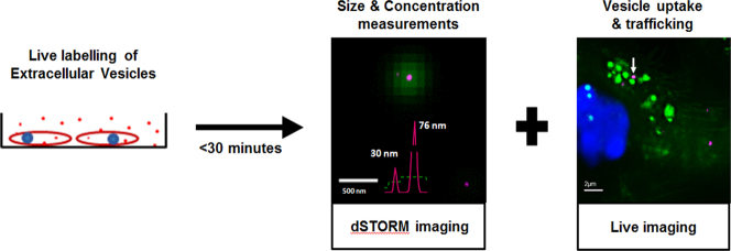
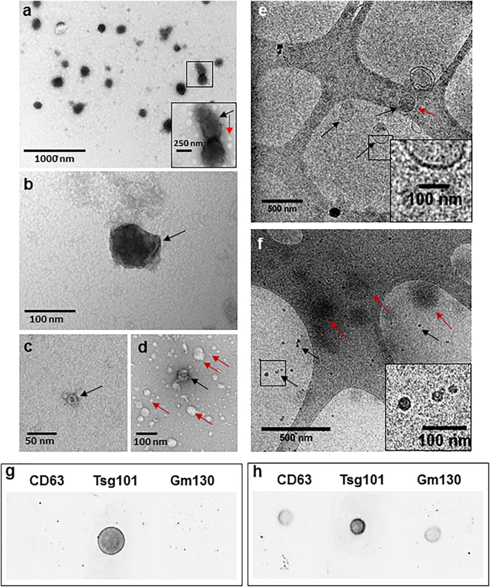
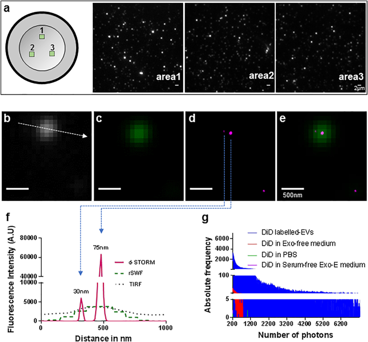
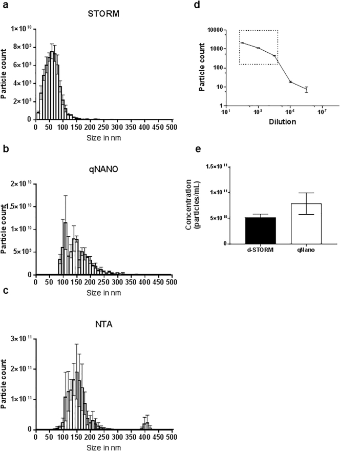
Extracellular vesicles (EVs) have prevalent roles in cancer biology and regenerative medicine. Conventional techniques for characterising EVs including electron microscopy (EM), nanoparticle tracking analysis (NTA) and tuneable resistive pulse sensing (TRPS), have been reported to produce high variability in particle count (EM) and poor sensitivity in detecting EVs below 50 nm in size (NTA and TRPS), making accurate and unbiased EV analysis technically challenging. This study introduces direct stochastic optical reconstruction microscopy (d-STORM) as an efficient and reliable characterisation approach for stem cell-derived EVs. Using a photo-switchable lipid dye, d-STORM imaging enabled rapid detection of EVs down to 20-30 nm in size with higher sensitivity and lower variability compared to EM, NTA and TRPS techniques. Imaging of EV uptake by live stem cells in culture further confirmed the potential of this approach for downstream cell biology applications and for the analysis of vesicle-based cell-cell communication.

Keywords: Extracellular vesicle; Stem cell; Super-resolution microscopy.


Figures

Unlabelled Image
Graphical abstract
Fig. 1
Fig. 1
Characterisation of EVs isolated from MSC culture supernatant. (a–d) TEM images of EVs. (a) TEM showing polydispersity EV structures of 100–250 nm EVs. Zoomed-in box shows cup-shaped morphology of a vesicle (black arrow) and non-cup-shaped white vesicular structures below 50 nm in size (red arrow). (b) 100 nm vesicle, black arrow showing membrane fold. (c) 20 nm cup-shaped EV (black arrow). (d) 50 nm cup-shaped EV structure (black arrow) along with other white, non-cup-shaped vesicular structures of 5 to 100 nm in size (red arrows). (e and f) Cryo-TEM images of EVs. (e) Image showing spherical (black arrows) and elongated (red arrow) EV structure with a clear membrane (zoomed-in box). (f) Images showing 20 nm EVs with defined borders (black arrows and zoomed-in box) and larger EVs with undefined borders (red arrows). Scale bars are defined in each figure. (g–h) Dot blot immunodetection of CD63, TSG101 and GM130 proteins in (g) EV lysate and (h) cell lysate samples isolated from MSCs.
Fig. 2
Fig. 2
d-STORM imaging of DiD-labelled EVs isolated from MSCs. (a) Graphical representation of the sample scanning pattern with snapshots of areas 1, 2 and 3 recorded in TIRF. Scale bar: 2 μm. (b–f) d-STORM images of MSC-derived EVs showing (b) TIRF (white), (c) rSWF (sum of wide field frames over the experiment) (green 75), (d) d-STORM (magenta) and (e) merge of rSWF and d-STORM. Scale bar: 500 nm. (f) Cross-sectional line-profiling of 2 EVs imaged in TIRF, rSWF and d-STORM as shown in (b) to (e), measured across the line indicated as dotted arrow in (b). (g) Absolute frequency of photon number recorded in [DiD in PBS] (green), [DiD in Exo-E medium] (red), [DiD in serum-free Exo-E medium] (purple) and DiD-labelled EV sample (blue).
Fig. 3
Fig. 3
Size distribution and concentration analysis of EVs isolated from MSCs. (a–c) Particle size distribution analysed by (a) d-STORM imaging, (b) TRPS and (c) NTA. (d) Standard curve from DiD-labelled EV dilutions to determine the working range for d-STORM particle count, shown in the boxed area. (e) EV concentration measurements produced by d-STORM and TRPS based approaches. Data represented as mean ± SEM, *p < 0.05 (n = 3).
Fig. 4
Fig. 4
Cellular uptake of MSC-derived EVs imaged in live NSC culture. (a–b) Confocal microscopy imaging of live DiO-labelled NSCs (green) with nuclear counterstain (blue), incubated in the (a) presence or (b) absence of DiD-labelled EVs. Arrows show DiD-labelled EVs in magnified view of boxed area. (c) SIM imaging of DiD-labelled EVs (red) uptake by DiO-labelled NSCs (green), with magnified areas 1 and 2 showing intracellular DiD-labelled EVs (arrows).
Supplementary Fig. 1
Image analysis of EVs captured using d-STORM super resolution microscopy. (a–c) Snapshots describing the precision of d-STORM gaussian plot. (a) Gaussian rendered image of a vesicular structure (white). (b) Cross plot (red) of localised molecules used to render gaussian image. (c) Merged image of gaussian and cross plot. (d–f) Snapshots describing the ImageJ analysis of exported d-STORM images. (d) Exported d-STORM image. (e) ImageJ macro mapping of vesicle shape and size. (f) ImageJ export of particle analysis. Yellow highlights point out the structures with circularity below 0.5.
Supplementary Fig. 2
Photo-blinking frequency of DiD-labelled EVs isolated from MSCs during d-STORM imaging. (a) Screenshot from the video used to calculate the photo-blinking frequency of DiD-labelled EVs during STORM imaging, showing a positive blinking region with EVs (blue circle) and a background region for blank reading (red circle). (b) Graphical representation of the photo-blinking frequency of DiD-labelled EVs (blue) against blank region (red).

References

    1. van Niel G., D'Angelo G., Raposo G. Shedding light on the cell biology of extracellular vesicles. Nat. Rev. Mol. Cell Biol. 2018;19(4):213. - PubMed
    1. Valadi H., Ekström K., Bossios A., Sjöstrand M., Lee J.J., Lötvall J.O. Exosome-mediated transfer of mRNAs and microRNAs is a novel mechanism of genetic exchange between cells. Nat. Cell Biol. 2007;9(6):654–659. - PubMed
    1. Katsuda T., Tsuchiya R., Kosaka N. Human adipose tissue-derived mesenchymal stem cells secrete functional neprilysin-bound exosomes. Sci. Rep. 2013;3(1197) - PMC - PubMed
    1. Wu J., Qu Z., Fei Z.-W., Wu J.-H., Jiang C.-P. Role of stem cell-derived exosomes in cancer. Oncol. Lett. 2017;13(5):2855–2866. - PMC - PubMed
    1. Chivet M., Hemming F., Pernet-Gallay K., Fraboulet S., Sadoul R. Emerging role of neuronal exosomes in the central nervous system. Front. Physiol. 2012;3(145) - PMC - PubMed

Publication types