

Three dimensional evaluation of soft tissue after orthognathic surgery
- PMID: 30290762
- PMCID: PMC6173856
- DOI: 10.1186/s13005-018-0179-z
Three dimensional evaluation of soft tissue after orthognathic surgery
Abstract
Background: To evaluate the nasolabial soft tissue change three-dimensionally after orthognathic surgery, using a structured light scanner.
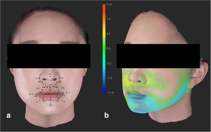
Methods: Thirty-two malocclusion patients, who underwent orthognathic surgery, were evaluated. CBCT and 3D facial scans were obtained before surgery and 3 months after surgery. The 3D changes in the 26 landmarks, and the relative ratio of the soft tissue movement to the bony movement, were evaluated.
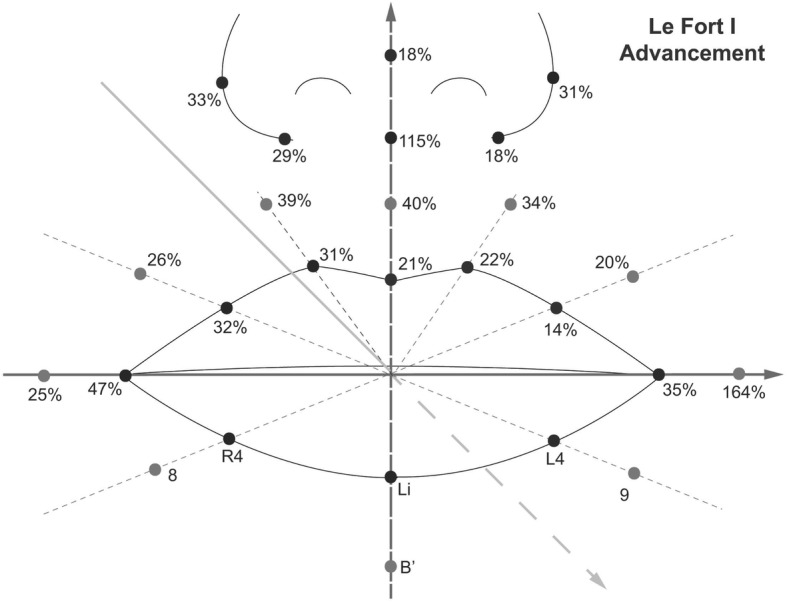
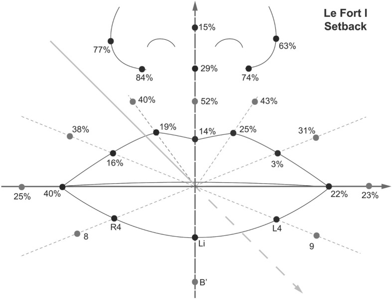
Results: In the Le Fort I advancement patients, the nasal tip moved 17% forward, compared to the maxillary bony movement, but the nasal prominence decreased 15%. The alar width increased 4 mm after the advancement, and the width decreased 4.7 mm after Le Fort I setback. The relative ratio of the soft tissue movement to the bony movement after bilateral sagittal split osteotomy was about 66% at the Li point in the anteroposterior direction, and it was 21% in the Le Fort I advancement and 14% in Le Fort I setback at the Ls point.
Conclusion: Alar cinch suturing may not be sufficient to overcome the effect of the maxilla advancement compressing the nasal complex. Alar width widening was prevented in Le Fort I setback. However, it is uncertain that the alar cinch suturing was solely responsible. The soft tissue around the mandible tends to accompany the bony movement more than the maxillary area. In addition, structured light scanning system proved to be a useful tool to evaluate the nasolabial soft tissue.
Keywords: 3D measurement; Nasolabial soft tissue; Orthognathic surgery; Structured light-based scanners.
Conflict of interest statement
Ethics approval and consent to participate
This study was approved by the Ethics Committee at the Kyung Hee University Dental Hospital (KHD IRB 1603–4).
Consent for publication
Not applicable.
Competing interests
The authors declare, that they have no competing interests.
Publisher’s Note
Springer Nature remains neutral with regard to jurisdictional claims in published maps and institutional affiliations.
Figures

References
-
- Bailey LJ, Collie FM, White RP., Jr Long-term soft tissue changes after orthognathic surgery. Int J Adult Orthodon Orthognath Surg. 1996;11:7–18. - PubMed
MeSH terms
LinkOut - more resources
Full Text Sources
