. 2018 Nov 30;293(48):18601-18612.
doi: 10.1074/jbc.RA118.005695. Epub 2018 Oct 5.

Mechanistic basis for the evolution of chalcone synthase catalytic cysteine reactivity in land plants

Affiliations

Mechanistic basis for the evolution of chalcone synthase catalytic cysteine reactivity in land plants

Geoffrey Liou et al. J Biol Chem. .

Abstract

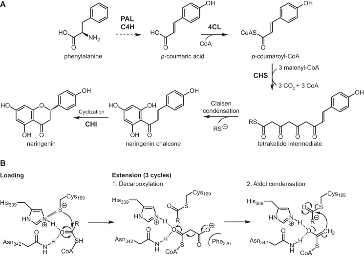
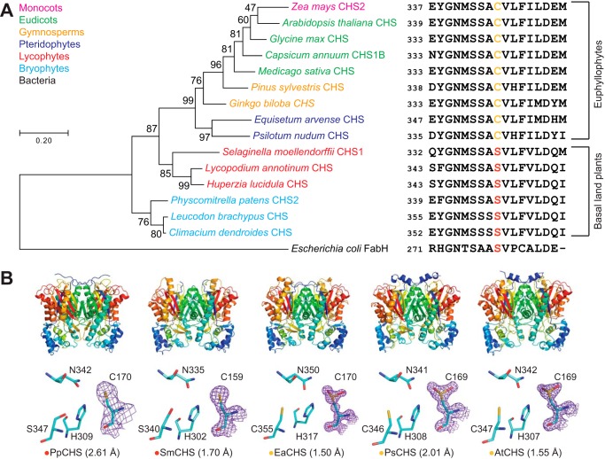
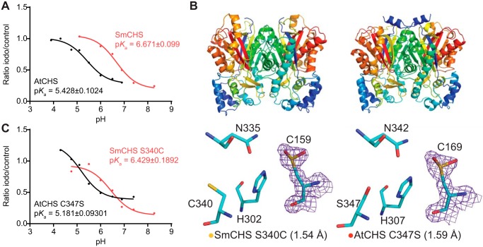
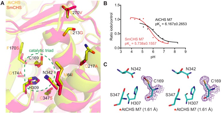
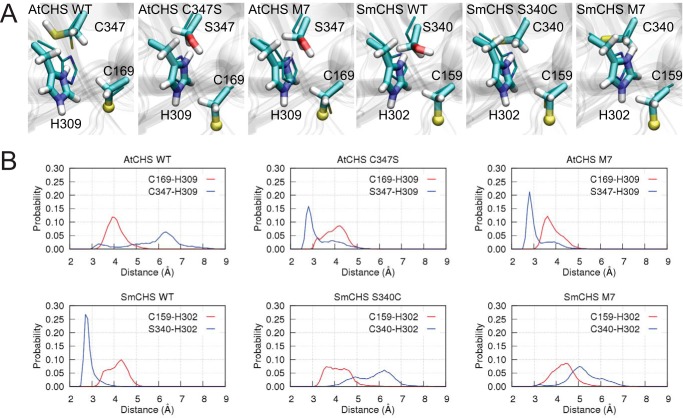
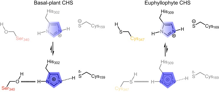
Flavonoids are important polyphenolic natural products, ubiquitous in land plants, that play diverse functions in plants' survival in their ecological niches, including UV protection, pigmentation for attracting pollinators, symbiotic nitrogen fixation, and defense against herbivores. Chalcone synthase (CHS) catalyzes the first committed step in plant flavonoid biosynthesis and is highly conserved in all land plants. In several previously reported crystal structures of CHSs from flowering plants, the catalytic cysteine is oxidized to sulfinic acid, indicating enhanced nucleophilicity in this residue associated with its increased susceptibility to oxidation. In this study, we report a set of new crystal structures of CHSs representing all five major lineages of land plants (bryophytes, lycophytes, monilophytes, gymnosperms, and angiosperms), spanning 500 million years of evolution. We reveal that the structures of CHS from a lycophyte and a moss species preserve the catalytic cysteine in a reduced state, in contrast to the cysteine sulfinic acid seen in all euphyllophyte CHS structures. In vivo complementation, in vitro biochemical and mutagenesis analyses, and molecular dynamics simulations identified a set of residues that differ between basal-plant and euphyllophyte CHSs and modulate catalytic cysteine reactivity. We propose that the CHS active-site environment has evolved in euphyllophytes to further enhance the nucleophilicity of the catalytic cysteine since the divergence of euphyllophytes from other vascular plant lineages 400 million years ago. These changes in CHS could have contributed to the diversification of flavonoid biosynthesis in euphyllophytes, which in turn contributed to their dominance in terrestrial ecosystems.

Keywords: chalcone synthase; cysteine; enzyme catalysis; enzyme mechanism; flavonoid; pKa; plant evolution; polyketide; polyketide synthase; redox regulation.


Conflict of interest statement

J. K. W. is a co-founder, a member of the Scientific Advisory Board, and a shareholder of DoubleRainbow Biosciences, which develops biotechnologies related to natural products

Figures

Figure 1.
Figure 1.
A, phenylpropanoid and flavonoid metabolism. PAL, phenylalanine ammonia-lyase; C4H, trans-cinnamate 4-monooxygenase; 4CL, 4-coumarate-CoA ligase; CHI, chalcone isomerase; CoA, coenzyme A. Cyclization of naringenin chalcone to naringenin also proceeds spontaneously in aqueous solution. B, reaction mechanism of CHS. The extension step is performed three times to repeatedly extend the starter molecule malonyl-CoA to form a linear tetraketide intermediate, which then cyclizes to form naringenin chalcone.
Figure 2.
Figure 2.
Structural and in vivo functional characterization of diverse CHS orthologs. A, maximum-likelihood phylogenetic tree of CHSs from diverse land-plant species, with clades indicated by color. The tree is rooted on a bacterial KAS III enzyme (EcFabH). The scale bar indicates evolutionary distance in substitutions per amino acid. The sequence near the differentially conserved cysteine/serine (position 347 in AtCHS) is shown for each CHS. B, overall apo crystal structures and active-site structures of CHSs from diverse plant lineages. Top, the homodimeric form of CHS is shown with a color gradient from blue at the N terminus to red at the C terminus of each monomer. Bottom, backbone and side chains of the catalytic triad and the differentially conserved cysteine/serine are shown. The 2FoFc electron density map contoured at 1.5σ is shown around the catalytic cysteine. CHSs from euphyllophytes show the catalytic cysteine oxidized to sulfinic acid, whereas CHSs from basal land plants have a reduced catalytic cysteine. The red or yellow dot next to the enzyme name indicates the presence of serine or cysteine, respectively, in position 347 (AtCHS numbering).
Figure 3.
Figure 3.
pKa measurement of the catalytic cysteine and characterization of key residues that affect pKa. A, pKa measurement of AtCHS and SmCHS WT enzymes. CHS enzyme was pre-incubated at various pH values with or without the 25 μm iodoacetamide inhibitor for 30 s, and an aliquot was taken to run in a CHS activity assay. The ratio of naringenin product produced in the iodoacetamide treatment divided by the control treatment was calculated for each pH point. A nonlinear regression was performed to fit a log(inhibitor) versus response curve to determine the pH at which 50% of maximal inhibition was achieved, which was determined to be the pKa value of the catalytic cysteine residue. The pKa of AtCHS is close to the 5.5 determined for other euphyllophyte CHSs, whereas the pKa of SmCHS is over 1 pH unit higher. B, overall structures and active-site configurations of AtCHS C347S and SmCHS S340C single mutants. The 2FoFc electron density map contoured at 1.5σ is shown around the catalytic cysteine. SmCHS S340C shows oxidation of Cys-159, unlike the SmCHS WT. AtCHS C347S has an oxidized Cys-169, like AtCHS WT. C, pKa measurements of AtCHS C347S and SmCHS S340C mutants.
Figure 4.
Figure 4.
Identification and characterization of additional key residues that affect CHS cysteine reactivity. A, overlaid crystal structures of AtCHS and SmCHS showing the seven conserved residue differences between euphyllophyte and basal-plant CHSs. B, pKa measurement of AtCHS M7 and SmCHS M7 mutants. The pKa of each M7 mutant is about 0.5 pH units higher or lower, respectively, than the corresponding WT CHS. C, active sites of the two monomers of the AtCHS M7 septuple mutant structure. The 2FoFc electron density map contoured at 1.5σ is shown around the catalytic cysteine. The crystal structure shows oxidation to sulfenic acid in the catalytic cysteine of one chain (left) and a reduced cysteine in the other (right).
Figure 5.
Figure 5.
Molecular dynamics simulations of CHS orthologs and mutants. A, centroid structure of the largest cluster of the catalytic pair Cys-169–His-309. For visualization purposes, the sulfur atom in the ionic cysteine Cys-169 is shown in a ball representation, and crystal structures are depicted as thin sticks. B, distributions of inter-residue distances obtained from simulations.
Figure 6.
Figure 6.
Proposed model for differential modulation of catalytic cysteine nucleophilicity in basal-plant (left) and euphyllophyte (right) CHSs. In basal-plant CHSs (left), the serine (Ser-340 in SmCHS) interacts more strongly with the histidine of the catalytic triad, weakening the ionic interaction that stabilizes the thiolate form of the catalytic cysteine. This is depicted as a shift of the equilibrium toward a state in which the positive charge on the histidine (blue) is shifted away from the catalytic cysteine (Cys-159 in SmCHS), and the shared proton interacts more closely with cysteine. In euphyllophyte CHSs (right), this position mutated to a cysteine (Cya-347 in AtCHS), which interacts relatively loosely with the catalytic histidine, in turn strengthening the ionic interaction between the catalytic histidine and the activated thiolate of the catalytic cysteine. This is depicted as a shift of the equilibrium toward a state in which the positive charge on the histidine (blue) is shifted toward the catalytic cysteine (Cys-169 in AtCHS).

References

    1. Weng J.-K., and Chapple C. (2010) The origin and evolution of lignin biosynthesis. New Phytol. 187, 273–285 10.1111/j.1469-8137.2010.03327.x - DOI - PubMed
    1. Winkel-Shirley B. (2001) Flavonoid biosynthesis. A colorful model for genetics, biochemistry, cell biology, and biotechnology. Plant Physiol. 126, 485–493 10.1104/pp.126.2.485 - DOI - PMC - PubMed
    1. Yao L. H., Jiang Y. M., Shi J., Tomás-Barberán F. A., Datta N., Singanusong R., and Chen S. S. (2004) Flavonoids in food and their health benefits. Plant Foods Hum. Nutr. 59, 113–122 10.1007/s11130-004-0049-7 - DOI - PubMed
    1. Rausher M. D. (2006) in The Science of Flavonoids (Grotewold E., ed) pp. 175–211, Springer, New York
    1. Weng J.-K., and Noel J. P. (2013) Chemodiversity in Selaginella: a reference system for parallel and convergent metabolic evolution in terrestrial plants. Front. Plant Sci. 4, 119 - PMC - PubMed

Publication types

LinkOut - more resources