. 2018 Dec 7;293(49):18890-18902.
doi: 10.1074/jbc.RA118.004462. Epub 2018 Oct 5.

A dominant dendrite phenotype caused by the disease-associated G253D mutation in doublecortin (DCX) is not due to its endocytosis defect

Affiliations

A dominant dendrite phenotype caused by the disease-associated G253D mutation in doublecortin (DCX) is not due to its endocytosis defect

Chan Choo Yap et al. J Biol Chem. .

Abstract

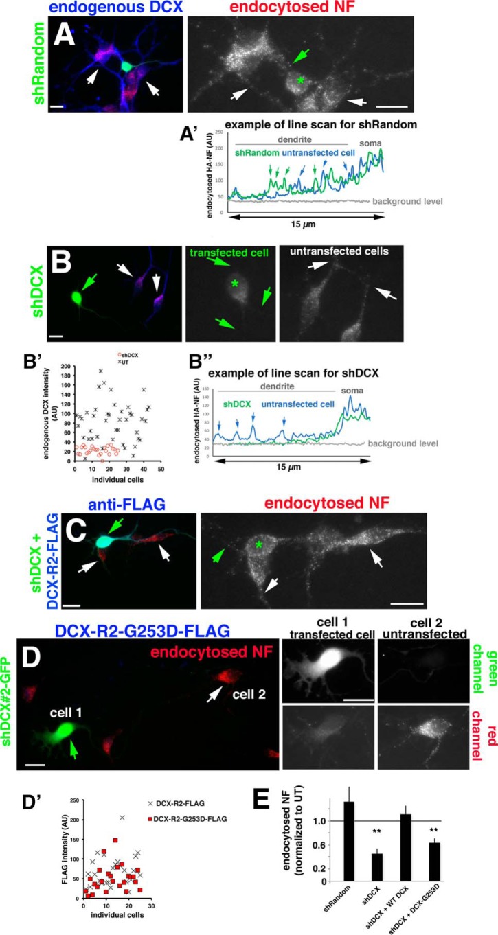
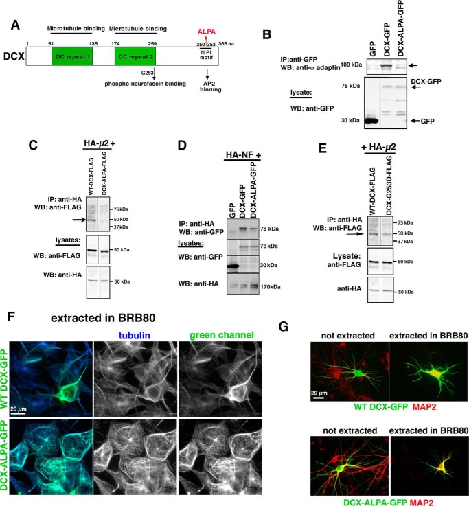
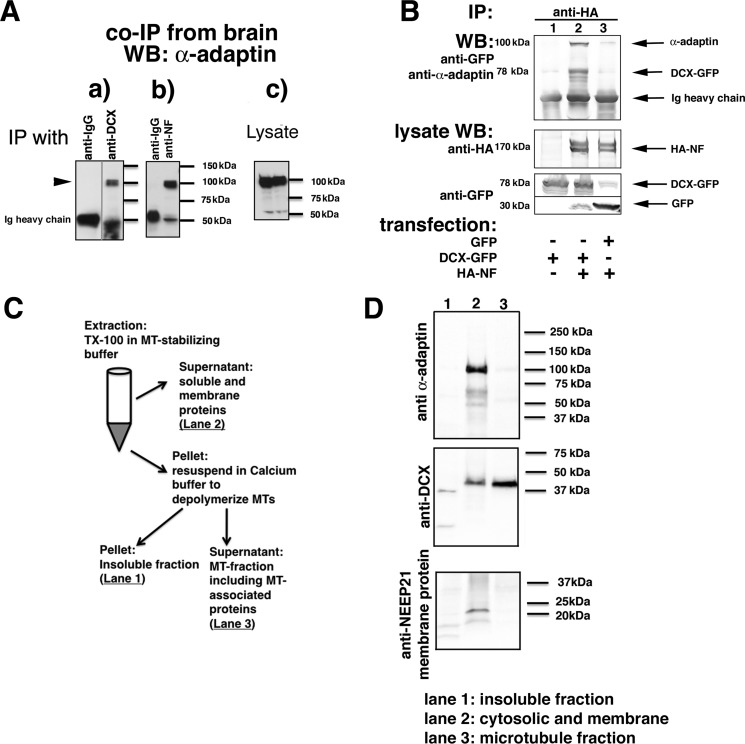
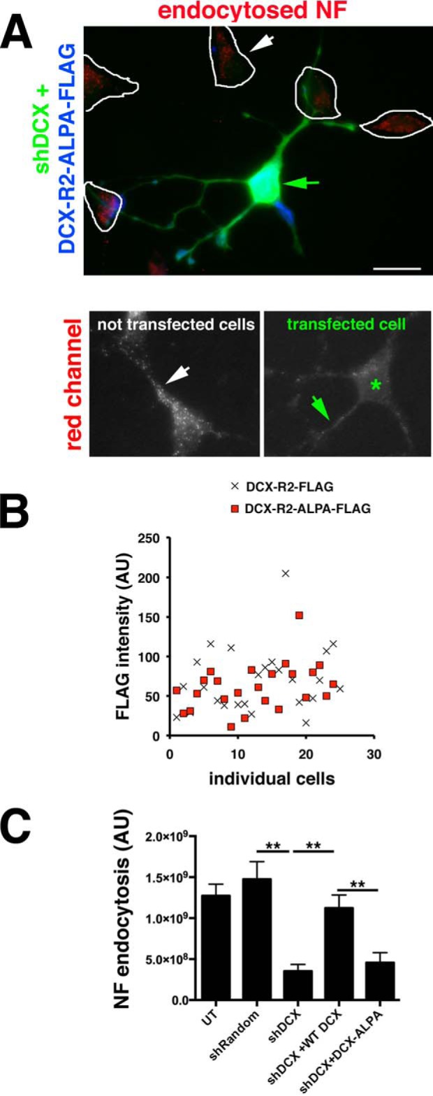
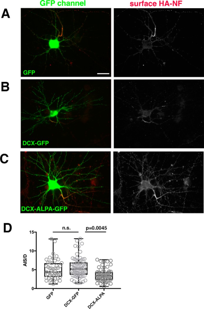
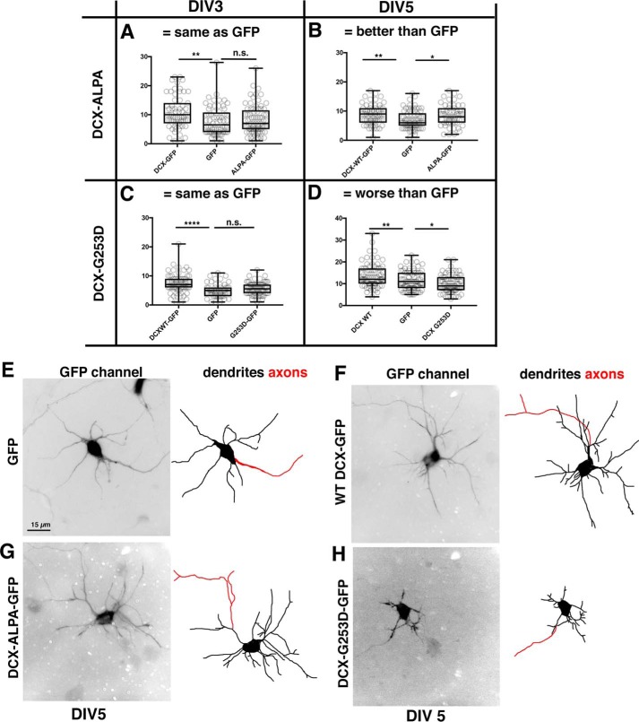
Doublecortin (DCX) is a protein needed for cortical development, and DCX mutations cause cortical malformations in humans. The microtubule-binding activity of DCX is well-described and is important for its function, such as supporting neuronal migration and dendrite growth during development. Previous work showed that microtubule binding is not sufficient for DCX-mediated promotion of dendrite growth and that domains in DCX's C terminus are also required. The more C-terminal regions of DCX bind several other proteins, including the adhesion receptor neurofascin and clathrin adaptors. We recently identified a role for DCX in endocytosis of neurofascin. The disease-associated DCX-G253D mutant protein is known to be deficient in binding neurofascin, and we now asked if disruption of neurofascin endocytosis underlies the DCX-G253D-associated pathology. We first demonstrated that DCX functions in endocytosis as a complex with both the clathrin adaptor AP-2 and neurofascin: disrupting either clathrin adaptor binding (DCX-ALPA) or neurofascin binding (DCX-G253D) decreased neurofascin endocytosis in primary neurons. We then investigated a known function for DCX, namely, increasing dendrite growth in cultured neurons. Surprisingly, we found that the DCX-ALPA and DCX-G253D mutants yield distinct dendrite phenotypes. Unlike DCX-ALPA, DCX-G253D caused a dominant-negative dendrite growth phenotype. The endocytosis defect of DCX-G253D thus was separable from its detrimental effects on dendrite growth. We recently identified Dcx-R59H as a dominant allele and can now classify Dcx-G253D as a second Dcx allele that acts dominantly to cause pathology, but does so via a different mechanism.

Keywords: axon initial segment; clathrin; clathrin adaptor; dendrite; dominant allele of DCX; endocytosis; lissencephaly; microtubule; multifunctional protein; neurite outgrowth; neurofascin; neuron.


Conflict of interest statement

The authors declare that they have no conflicts of interest with the contents of this article

Figures

Figure 1.
Figure 1.
DCX-G253D fails to rescue NF endocytosis in primary neurons after DCX knockdown. A–D, endocytosis of endogenous NF (red) was determined in DIV3 hippocampal neurons expressing shRandom-GFP (A), shDcx#2-GFP (B), shDcx#2-GFP plus rescue plasmid WT DCX-R2-FLAG (blue) (C), and shDcx#2-GFP plus rescue plasmid DCX-R2-G253D-FLAG (blue) (D). Enlarged views of the red channel (endocytosed NF) are shown on the right in A–C. Green star, transfected cell; arrows in A–C, dendrites. An example of the brightness of NF-containing endosomes is shown as line scans for shRandom (A′) and shDcx (B″) for transfected (green line) and untransfected cells (blue line). Arrows, peaks corresponding to endosomes along the dendrite. B′, levels of endogenous DCX were plotted for untransfected cells (UT; black crosses) and cells expressing shDcx#2-GFP (red circles). For D, cell 1 (green arrows) corresponds to a transfected cell, and cell 2 (white arrows) corresponds to an untransfected cell. Corresponding green and red channels are shown on the right. Scale bars, 20 μm. D′, DCX-R2-FLAG (cross) and DCX-R2-G253D-FLAG (red square) are expressed at comparable levels in transfected neurons. n = 25 cells for each plasmid. Transfected cultures were also counterstained with antibody against DCX to evaluate the extent of overexpression compared with untransfected cells (Fig. S1). E, quantification of NF endocytosis levels for conditions shown in A–D. Quantification of one representative experiment is shown. Measurements are normalized to untransfected (UT) cells in the same field. n = 15–25 cells/condition/experiment. Error bars, S.E. **, p < 0.001 (ANOVA with post hoc test).
Figure 2.
Figure 2.
DCX-ALPA is an AP-2–binding mutant. A, diagram of DCX domains indicating microtubule-binding (green boxes), phosphoneurofascin-binding (containing residue Gly-253), and AP-2–binding sites (YLPL at positions 350–353). 350YLPL353 was mutated to 350ALPA353 (ALPA). B–D, DCX-ALPA is deficient in binding AP-2 complex but still binds to HA-NF. HEK293 cells were transfected with the indicated plasmids, and immunoprecipitations were carried out as indicated. Immunoprecipitates were analyzed by Western blotting (WB) with the indicated antibodies. The lysate (5% of total) was probed by Western blotting as labeled. Arrows, the respective bands. All immunoprecipitations were carried out at least three times from independent lysates. E, DCX-G253D still binds to AP-2 complex. HEK293 cells were transfected with HA-μ2 together with WT DCX-FLAG (lane 1) or DCX-G253D-FLAG (lane 2). Immunoprecipitations (IP) were carried out with anti-HA antibody. Western blots were carried out with anti-FLAG antibody (top). Western blots of lysates are shown below. This immunoprecipitation was carried out at least two times from independent lysates. The blot in E represents additional lanes from the same experiment as C. The WT DCX lane is thus reused in E. Intervening irrelevant lanes were removed in B, C, and E. F, DCX-ALPA-GFP (green) remains stably associated with microtubules (blue, anti-tubulin) after detergent extraction in MT-stabilizing buffer BRB80. The panels on the right show each channel separately as well as a merged image. Scale bar, 20 μm. G, DCX-ALPA-GFP (green) is stably associated with microtubules in neurons after extraction in BRB80. MAP2 is counterstained in red.
Figure 3.
Figure 3.
A complex of NF, DCX, and AP-2 can be immunoprecipitated from transfected cells and brain. A, α-adaptin is immunoprecipitated with anti-DCX antibodies from embryonic rat brain membrane fractions (a) as well as by anti-NF antibodies (b). Nonimmune IgG-coated beads were used as negative controls. Levels of endogenous α-adaptin are shown in the lysate blots (c). An intervening irrelevant lane was removed in a. B, α-adaptin is only co-immunoprecipitated with HA-NF if DCX-GFP is co-expressed. HEK293 cells were transfected with the indicated plasmids, and immunoprecipitations (IP) were carried out as labeled. Immunoprecipitates were analyzed by Western blotting (WB) with the indicated antibodies. The lysate (5% of total) was probed by Western blotting as labeled. C and D, freshly dissociated E18 rat cortex was homogenized and fractionated according to the diagram (shown in C) into a soluble (cytosolic and membrane proteins), MT-associated, and insoluble fraction. The insoluble (lane 1), soluble/membrane (lane 2), and microtubule-associated (lane 3) fractions were separated by SDS-PAGE and blotted against α-adaptin, DCX, and a membrane protein (NEEP21).
Figure 4.
Figure 4.
DCX-ALPA does not rescue the NF endocytosis defect caused by down-regulation of DCX. A, endocytosis of endogenous NF (red) was determined in DIV3 hippocampal neurons expressing shDcx#2-GFP plus rescue plasmid Dcx-R2-ALPA-FLAG (blue). Enlarged views of the red channel are shown below. Green star, transfected cell; arrows, dendrites. Controls are the same as in Fig. 1 and are not shown again. Scale bar, 20 μm. B, DCX-R2-FLAG (cross) and DCX-R2-ALPA-FLAG (red square) are expressed at comparable levels in transfected neurons. n = 25 cells for each plasmid. Transfected cultures were also counterstained with antibody against DCX to compare the extent of overexpression compared with untransfected cell (Fig. S1). C, quantification of NF endocytosis levels. n = 25 cells/condition/experiment. One representative experiment is shown. Error bars, S.E. **, p < 0.001 (ANOVA with post hoc test).
Figure 5.
Figure 5.
Co-expression of DCX-ALPA interferes with HA-NF accumulation on the AIS. A–C, HA-NF (red) accumulates predominantly at the surface of the AIS in cells expressing GFP as a control (A) or DCX-GFP (B), but HA-NF mislocalizes to the somatic and dendritic surface when DCX-ALPA-GFP is expressed (C). The right panels show single-channel images of the HA-NF surface distributions (DIV9 neurons 20 h post-transfection). Scale bar, 20 μm. Transfected cultures were also counterstained with antibody against DCX to compare the extent of overexpression compared with control cells expressing GFP (Fig. S1). D, surface distribution of HA-NF was quantified by determining the surface intensity of HA-NF on the AIS versus dendrites (AIS/D PI) for neurons expressing GFP, DCX-GFP, or DCX-ALPA-GFP. ***, p < 0.0001; n.s., not significant. One representative experiment is shown (of a total of three independent experiments). n = 51 cells for GFP, 70 cells for DCX-GFP, and 59 cells for DCX-ALPA. Whisker plots show the median as a line, 25–75% as a box, and the data range as whiskers (Kruskal–Wallis test; p = 0.0045 for DCX-GFP versus DCX-ALPA; p = 0.0014 for GFP versus DCX-ALPA).
Figure 6.
Figure 6.
Divergent phenotypes of DCX-ALPA and DCX-G253D on dendrite growth. A–D, number of dendrites at DIV3 and DIV5 of at least 15-μm length from center of soma was determined for neurons expressing GFP, WT DCX-GFP, or DCX-ALPA-GFP (A and B) or DCX-G253D-GFP (C and D) for DIV3 (A and C) and DIV5 (B and D). One representative experiment is shown (of a total of three independent experiments). n = 50–75 cells/condition in each experiment. The axon was excluded from counts. Transfected cultures were also counterstained with antibody against DCX to compare the extent of overexpression compared with control cells expressing GFP (Fig. S1). For parametric data, we used one-way ANOVA followed by Fisher's least significant difference test. For nonparametric data sets, we used Kruskal–Wallis followed by uncorrected Dunn's multiple-comparison test. Whisker plots show the median as a line, 25–75% as a box, and the data range as whiskers. *, p < 0.05; **, p < 0.001; ****, p < 0.00001; n.s., not significant. Measurements from individual cells are also shown. E–H, representative examples of DIV5 neurons transfected with GFP (E), WT DCX (F), DCX-ALPA (G), or DCX-G253D (H). Dendrites were traced in the right-hand panels in black. The axon is traced in red.

References

    1. Liu J. S. (2011) Molecular genetics of neuronal migration disorders. Curr. Neurol. Neurosci. Rep. 11, 171–178 10.1007/s11910-010-0176-5 - DOI - PubMed
    1. Friocourt G., Marcorelles P., Saugier-Veber P., Quille M.-L., Marret S., and Laquerrière A. (2011) Role of cytoskeletal abnormalities in the neuropathology and pathophysiology of type I lissencephaly. Acta Neuropathol. 121, 149–170 10.1007/s00401-010-0768-9 - DOI - PMC - PubMed
    1. Dixon-Salazar T. J., and Gleeson J. G. (2010) Genetic regulation of human brain development: lessons from Mendelian diseases. Ann. N.Y. Acad. Sci. 1214, 156–167 10.1111/j.1749-6632.2010.05819.x - DOI - PMC - PubMed
    1. LoTurco J. J., and Bai J. (2006) The multipolar stage and disruptions in neuronal migration. Trends Neurosci. 29, 407–413 10.1016/j.tins.2006.05.006 - DOI - PubMed
    1. Bai J., Ramos R. L., Ackman J. B., Thomas A. M., Lee R. V., LoTurco J. J. (2003) RNAi reveals doublecortin is required for radial migration in rat neocortex. Nat. Neurosci. 6, 1277–1283 10.1038/nn1153 - DOI - PubMed

Publication types

MeSH terms