Review
. 2019 Oct 1;9(10):a033506.
doi: 10.1101/cshperspect.a033506.

Function and Dysfunction of TMC Channels in Inner Ear Hair Cells

Affiliations
Review

Function and Dysfunction of TMC Channels in Inner Ear Hair Cells

David P Corey et al. Cold Spring Harb Perspect Med. .

Abstract

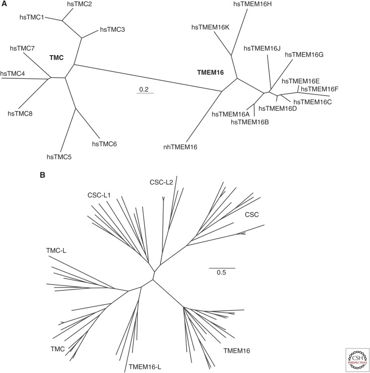
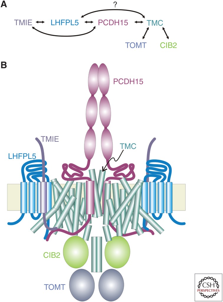
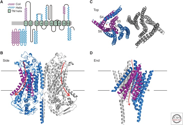
The TMC1 channel was identified as a protein essential for hearing in mouse and human, and recognized as one of a family of eight such proteins in mammals. The TMC family is part of a superfamily of seven branches, which includes the TMEM16s. Vertebrate hair cells express both TMC1 and TMC2. They are located at the tips of stereocilia and are required for hair cell mechanotransduction. TMC1 assembles as a dimer and its similarity to the TMEM16s has enabled a predicted tertiary structure with an ion conduction pore in each subunit of the dimer. Cysteine mutagenesis of the pore supports the role of TMC1 and TMC2 as the core channel proteins of a larger mechanotransduction complex that includes PCDH15 and LHFPL5, and perhaps TMIE, CIB2 and others.


Figures

Figure 1.
Figure 1.
Phylogeny of the TMC and TMEM16 families. (A) An unrooted phylogeny tree of the TMC and TMEM16 families, derived from an alignment of vertebrate TMCs and mammalian TMEM16s. Only Nectria and human orthologs are shown here. Within the TMC branch, TMC1–3 cluster in a distinct subfamily. (B) The TMC/TMEM16 superfamily. (Panel B based on data in Medrano-Soto et al. 2018.)
Figure 2.
Figure 2.
Possible schematic of the hair cell mechanotransduction apparatus. (A) Reported interactions among six proteins essential for mechanosensory transduction in hair cells. (B) A possible arrangement of proteins within the transduction apparatus. TMC1 and PCDH15 are both dimers, so it is attractive to suppose that there is a one-to-one relationship between them, with one PCDH15 opening the pore in one TMC subunit of the dimeric channel. In addition, PCDH15 and LHFPL5 can form a tetrameric complex (Ge et al. 2018) so LHFPL5 may also be near the dimer interface, rather than as depicted. There is little information about stoichiometries for the other proteins of the complex, nor is it known which are part of a mature complex and which are only necessary for assembly.
Figure 3.
Figure 3.
Predicted secondary and tertiary structure of TMC1. (A) Predicted secondary structure of TMC1. Transmembrane domains assigned by I-TASSER and Phyre2 are based on amino acid similarity to TMEM16A and solved structures of mmTMEM16A and nhTMEM16. Helix and coiled-coil predictions from PSSpred (Yan et al. 2013). (Reprinted from Pan et al. 2018 with permission from the authors.) (B) Tertiary structure of the dimeric TMC1 predicted by I-TASSER. The two subunits are colored blue and gray; the view is from within the plane of the membrane. The S10 transmembrane domains form the dimer interface. Within the blue subunit, the predicted pore-forming helices, S4-S7, are purple. The intracellular and extracellular loops are not well conserved with TMEM16 and are not well modeled by I-TASSER. Arrows show the predicted permeation pathway in each subunit. (C) Predicted arrangement of transmembrane helices; view from outside the cell. The red dot indicates the general region of the predicted pore. (D) Predicted arrangement of transmembrane helices; view from the plane of the membrane. Transmembrane helices S4–S7 form a groove that lines the pore. Most mutations in pore-facing residues of S4–S7 affect amplitude, single-channel conductance, or selectivity of the transduction current (Pan et al. 2018).

References

    1. Ahmed ZM, Goodyear R, Riazuddin S, Lagziel A, Legan PK, Behra M, Burgess SM, Lilley KS, Wilcox ER, Riazuddin S, et al. 2006. The tip-link antigen, a protein associated with the transduction complex of sensory hair cells, is protocadherin-15. J Neurosci 26: 7022–7034. - PMC - PubMed
    1. Ahmed ZM, Masmoudi S, Kalay E, Belyantseva IA, Mosrati MA, Collin RW, Riazuddin S, Hmani-Aifa M, Venselaar H, Kawar MN, et al. 2008. Mutations of LRTOMT, a fusion gene with alternative reading frames, cause nonsyndromic deafness in humans. Nat Genet 40: 1335–1340. - PMC - PubMed
    1. Alagramam KN, Murcia CL, Kwon HY, Pawlowski KS, Wright CG, Woychik RP. 2001a. The mouse Ames waltzer hearing-loss mutant is caused by mutation of Pcdh15, a novel protocadherin gene. Nat Genet 27: 99–102. - PubMed
    1. Alagramam KN, Yuan H, Kuehn MH, Murcia CL, Wayne S, Srisailpathy CR, Lowry RB, Knaus R, Van Laer L, Bernier FP, et al. 2001b. Mutations in the novel protocadherin PCDH15 cause Usher syndrome type 1F. Hum Mol Genet 10: 1709–1718. - PubMed
    1. Asai Y, Pan B, Nist-Lund C, Galvin A, Lukashkin AN, Lukashkina VA, Chen T, Zhou W, Zhu H, Russell IJ, et al. 2018. Transgenic Tmc2 expression preserves inner ear hair cells and vestibular function in mice lacking Tmc1. Sci Rep 8: 12124. - PMC - PubMed

Publication types

Substances