. 2018 Oct 5;8(1):14891.
doi: 10.1038/s41598-018-33143-1.

Structural insights into pro-aggregation effects of C. elegans CRAM-1 and its human ortholog SERF2

Affiliations

Structural insights into pro-aggregation effects of C. elegans CRAM-1 and its human ortholog SERF2

Meenakshisundaram Balasubramaniam et al. Sci Rep. .

Abstract

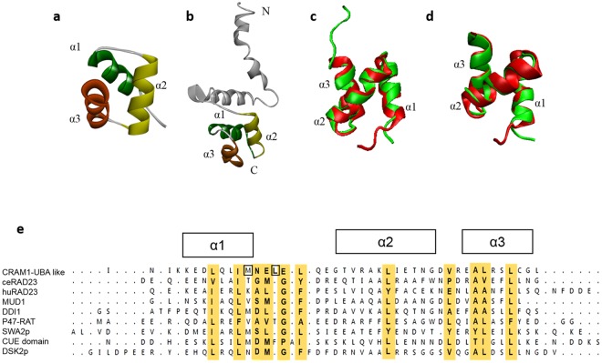
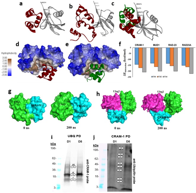
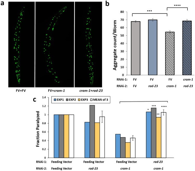
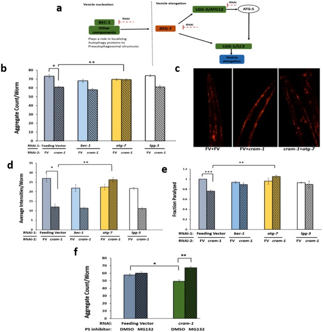
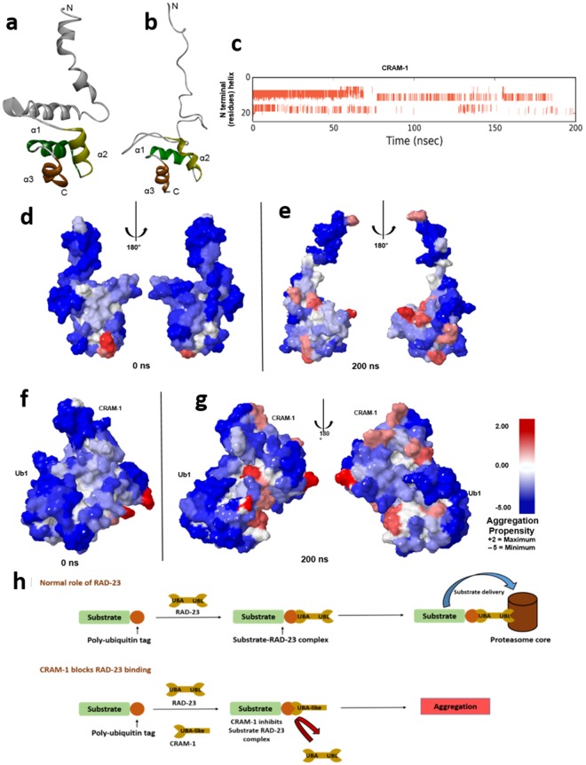
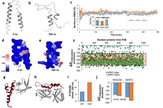
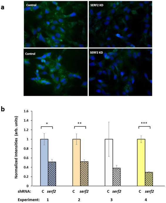
Toxic protein aggregates are key features of progressive neurodegenerative diseases. In addition to "seed" proteins diagnostic for each neuropathy (e.g., Aβ1-42 and tau in Alzheimer's disease), aggregates contain numerous other proteins, many of which are common to aggregates from diverse diseases. We reported that CRAM-1, discovered in insoluble aggregates of C. elegans expressing Q40::YFP, blocks proteasomal degradation of ubiquitinated proteins and thus promotes aggregation. We now show that CRAM-1 contains three α-helical segments forming a UBA-like domain, structurally similar to those of mammalian adaptor proteins (e.g. RAD23, SQSTM1/p62) that shuttle ubiquitinated cargos to proteasomes or autophagosomes for degradation. Molecular modeling indicates that CRAM-1, through this UBA-like domain, can form tight complexes with mono- and di-ubiquitin and may thus prevent tagged proteins from interacting with adaptor/shuttle proteins required for degradation. A human ortholog of CRAM-1, SERF2 (also largely disordered), promotes aggregation in SH-SY5Y-APPSw human neuroblastoma cells, since SERF2 knockdown protects these cells from amyloid formation. Atomistic molecular-dynamic simulations predict spontaneous unfolding of SERF2, and computational large-scale protein-protein interactions predict its stable binding to ubiquitins. SERF2 is also predicted to bind to most proteins screened at random, although with lower average stability than to ubiquitins, suggesting roles in aggregation initiation and/or progression.


Conflict of interest statement

The authors declare no competing interests.

Figures

Figure 1
Figure 1
Predicted 3-dimensional structure of CRAM-1 indicates a UBA-like domain. Structures of the CRAM-1 C-terminal region (a) and the full-length model (b) show the same bundle of three helices connected by loops. (c) Structural comparison of the CRAM-1 C-terminal region (red) with the UBA domain of human RAD23A (green). Superimposed structures illustrate structural agreement, with RMSD (root-mean-square deviation) = 1.14 Å. (d) C-terminal region of CRAM-1 (red) superimposed on the UBA domain of ceRAD-23 (green), RMSD = 1.02 Å. (e) Sequence alignment, based on superimposition of 3-dimensional structures, showing 3 α helices with conserved hydrophobic residues (highlighted in gold) and nearby exposed hydrophobic residues (boxes).
Figure 2
Figure 2
CRAM-1 and RAD-23 have similar interactions with ubiquitins. (a) The CRAM-1 UBA-like domain (red) interacts with Ub1 (mono-ubiquitin, gray); interacting amino acids of ubiquitin are labeled. (b) The full-length structure of CRAM-1 (red) binds the same region of ubiquitin, contacting most of the same ubiquitin residues that were predicted to interact with the CRAM-1 C-terminus alone. (c) Structural superimposition of ceRAD-23 (green) and CRAM-1 (red), showing their predicted interactions with mono-ubiquitin via the same ubiquitin aspects. (d,e) Binding of CRAM-1 (d, red) or ceRAD-23 (e, green) ribbon models to di-ubiquitin (space-filling model) is facilitated by hydrophobic interactions (brown; see scale at left). (f) Predicted interaction energies (ΔEinteraction) for CRAM-1 (UBA-like) and 3 other UBA proteins, each with mono-, di-, and tetra-ubiquitin. (g,h) Simulated structures of complexes between the CRAM-1 UBA domain (cyan) and either mono-ubiquitin (light green; g) or di-ubiquitin (pink and light green; h) appear stable over a 200-ns simulation. (i,j) Western blot analyses for CRAM-1 interaction to ubiquitin: lysates from wild-type N2 worms at days 1 and 5 were immuno-precipitated with biotinylated CRAM-1 antibody (i) or antibody to ubiquitin (j), resolved in polyacrylamide gradient gel lanes (10% w/v, BioRad), electroblotted to nylon membranes, and probed with antibody to CRAM-1 (i) or ubiquitin (j), followed by peroxidase-tagged antibody to IgG and chemiluminescence imaging (Western ECL kit, Bio-Rad).
Figure 3
Figure 3
Rescue by cram-1 knockdown, of both Q40::YFP and Aβ1–42 amyloid aggregation, requires RAD23. (a,b) Protection from Q40::YFP aggregation, conferred by cram-1 knockdown, is blocked by rad-23 RNAi. Dual knockdowns were conducted as described. Aggregate numbers per worm, ± SEM, were counted at day 5 post-hatch (D5PH) for 10–16 worms per group. Significance (2-tailed t-tests): ***P < 3E–5 comparing [FV + FV] to [FV + cram-1KD]; ****P < 10–4 comparing [FV + cram-1KD] to [rad-23KD  + cram-1KD]. (c) CL4176 worms were treated, beginning 36–40 h after egg isolation, with dual RNAi in 3 experiments. Cram-1 RNAi reduced paralysis relative to [FV + FV] controls, but not when cram-1KD was paired with rad23KD. Shaded bars show fraction paralyzed, normalized to FV, for 3 independent experiments; open bars summarize combined data ± SEM. Significance by chi2 test within each experiment, comparing [cram-1KD  + FV] to [cram-1KD  + rad23KD], for 50–100 worms/group: *P < 0.05; **P < 0.01; ***P = 0.001. Treating each experiment as one point per group, ****P < 0.006 by 2-tailed paired t-test (white/open bars).
Figure 4
Figure 4
Cram-1 knockdown reduces aggregation of Q40::YFP and formation of Aβ1–42 amyloid, dependent on proteasome and ATG-7 functions. (a) Schematic depiction of RNAi targets and their roles in autophagy pathways. (b) For dual RNAi, AM141 worms were fed from the L3/L4 molt on bacteria carrying empty FV, or FV expressing dsRNAs to target lgg-3, bec-1, or atg-7, each mixed 1:1 with RNAi against cram-1 (striped bars, ± SEM) or empty FV (solid bars, ± SEM). Aggregates per worm were counted on D5PH. *P < 2E–05, in a 1-tailed t-test comparing FV to [cram-1KD + FV]; **P < 0.0002, in a 2-tailed t-test comparing [cram-1KD + FV] to [cram-1KD + atg-7KD]. (c,d) LN149 worms expressing mCherry::ubiquitin in muscle were fed from the L3/L4 molt through D8PH, on dual-RNAi as described for panel b. Images of mCherry fluorescence are shown in c, and mean mCherry intensity per worm ± SEM is summarized in d. *P < 1E–04, in 1-tailed t-test between FV and [cram-1KD + FV]; **P < 6E–05, for a 2-tailed t-test of [cram-1KD + FV] vs. [cram-1KD + atg-7KD]. (e) CL4176 worms, expressing human Aβ1–42 in muscle, were fed dual RNAi as in b. Paralysis, assessed 36–40 h after induction for 50–100 worms/group, is shown as mean ± SEM, treating each experiment as one data point per group. One replicate value for [lgg-3KD + cram-1KD] was excluded as an outlier, >8.5 SDs from the mean of all other values (P < 6E–10). **Significance by 1-tailed paired t-test, P < 0.01. ***P < 1E–40, assuming a normal distribution. (f) C. elegans strain AM141 (unc54p/Q40::yfp) was fed from hatch on bacteria containing feeding vector (FV) or FV expressing dsRNA to target cram-1. Worms were treated from the L3/L4 molt onward with either 20-μM MG132 to inhibit proteasomes (cross-hatched bars), or vehicle only (solid bars). Aggregates per worm were counted on D5PH for 10–15 worms/group. *P ≈ 0.003, 1-tailed t-test between FV and cram-1KD; **P ≈ 1E–05, 2-tailed t-test for [cram-1KD + DMSO] vs. [cram-1KD + MG132].
Figure 5
Figure 5
Atomistic molecular-dynamic simulation predicts aggregation-prone regions in CRAM-1 due to structural unfolding. (a,b) MD simulation in Desmond (Desmond Molecular Dynamics System, ver. 2016.4, D.E. Shaw Research, New York, NY) showing the unfolding of protein structure from initial (a) to simulation-stability (b) conformations. (c) Helical regions (red bars) indicate unfolding of N-terminal residues, plotted against simulation time (x axis). (dg) Predicted aggregation propensity (see scale at right) of CRAM-1 alone before simulation of structural rearrangement (d), or at the end of simulation (e) and of CRAM-1 bound to mono-ubiquitin (ub1) before simulation (f), or at the end of simulation (g). (h) Schematic depiction of a proposed mechanism, wherein CRAM-1 competes with RAD-23 for binding to ubiquitin, thus impeding clearance of ubiquitinated substrates.
Figure 6
Figure 6
MD simulation and protein-protein interactions predict that SERF2 is aggregation-prone. (a,b) MD simulation of SERF2, in Desmond (Desmond Molecular Dynamics System, ver. 2016.4, Shaw Research, New York), indicating its initial state (a) and the unfolded state after 500 ns (b). (c) RMSD for three independent simulations of SERF2 structure monitored throughout the simulation. The inset shows the coefficient of variation for RMSD in each simulation. (d,e) Predicted aggregation-prone regions (see scale at left) for SERF2 in its initial conformation (d) and unfolded conformation after 500 ns of simulation (e). (f) Interaction energies were predicted for SERF2 in its initial (black dots) and simulated unfolded (green dots) conformations when interacting with 1000 random proteins from the PDB databank. Interaction energies are also shown for SERF2 dimer in its initial conformation (boxed red dot) and in a subsequent unfolded state (boxed yellow dot). The tinted rectangle indicates the 90% confidence interval for SERF2 interaction energies. (g,h) Predicted structures are shown for SERF2 interacting with Ub1 (g) or Ub2 (h). (i) Average H-bond number was calculated for a single SERF2 molecule interacting with Ub1 or Ub2, over a 200-ns simulation. (j) Interaction energies were predicted for RAD23A or SERF2 molecules binding to mono- and di-ubiquitin (Ub1 and Ub2, respectively).
Figure 7
Figure 7
SERF2 knockdown protects against amyloid aggregation in human neural cells. (a) SH-SY5Y-APPSw neuroblastoma cells were transfected with SERF2 shRNA, and stained with thioflavin T at 48 h post-transfection. Amyloid fluorescence is displayed in green, and nuclei (counterstained with DAPI) appear blue. (b) Thioflavin-T fluorescence was quantified and divided by the number of DAPI-stained nuclei per field, to estimate amyloid deposition per cell. In 4 independent experiments, SERF-2 knockdown reduced amyloid by 50–70%, attaining statistical significance in 3 of 4 experiments. Fluorescence intensity per cell is plotted ± SEM, normalized to the control mean for each experiment. *P < 0.01 by 2-tailed t-test (initial experiment); **P < 0.01 by 1-tailed t-test; ***P < 0.001 by 1-tailed t-test.

Similar articles

Cited by

References

    1. Ayyadevara S, et al. Proteins in aggregates functionally impact multiple neurodegenerative disease models by forming proteasome-blocking complexes. Aging Cell. 2015;14:35–48. doi: 10.1111/acel.12296. - DOI - PMC - PubMed
    1. Kopito RR, Ron D. Conformational disease. Nat. Cell Biol. 2000;2:E207–E209. doi: 10.1038/35041139. - DOI - PubMed
    1. Stefani M, Dobson CM. Protein aggregation and aggregate toxicity: new insights into protein folding, misfolding diseases and biological evolution. J. Mol. Med. (Berl) 2003;81:678–699. doi: 10.1007/s00109-003-0464-5. - DOI - PubMed
    1. Youmans KL, et al. Intraneuronal Abeta detection in 5xFAD mice by a new Abeta-specific antibody. Mol. Neurodegener. 2012;7:8. doi: 10.1186/1750-1326-7-8. - DOI - PMC - PubMed
    1. Kikis EA, Gidalevitz T, Morimoto RI. Protein homeostasis in models of aging and age-related conformational disease. Adv. Exp. Med. Biol. 2010;694:138–159. doi: 10.1007/978-1-4419-7002-2_11. - DOI - PMC - PubMed

Publication types

MeSH terms

LinkOut - more resources