. 2018 Oct 5;8(1):14863.
doi: 10.1038/s41598-018-33286-1.

Fiber-bundle illumination: realizing high-degree time-multiplexed multifocal multiphoton microscopy with simplicity

Affiliations

Fiber-bundle illumination: realizing high-degree time-multiplexed multifocal multiphoton microscopy with simplicity

Jiun-Yann Yu et al. Sci Rep. .

Abstract

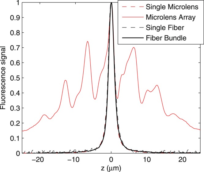
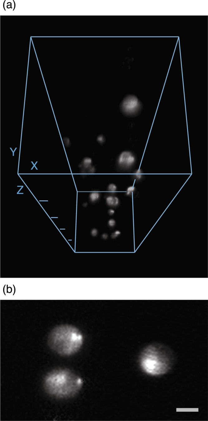
High-degree time-multiplexed multifocal multiphoton microscopy was expected to provide a facile path to scanningless optical-sectioning and the fast imaging of dynamic three-dimensional biological systems. However, physical constraints on typical time multiplexing devices, arising from diffraction in the free-space propagation of light waves, lead to significant manufacturing difficulties and have prevented the experimental realization of high-degree time multiplexing. To resolve this issue, we have developed a novel method using optical fiber bundles of various lengths to confine the diffraction of propagating light waves and to create a time multiplexing effect. Through this method, we experimentally demonstrate the highest degree of time multiplexing ever achieved in multifocal multiphoton microscopy (~50 times larger than conventional approaches), and hence the potential of using simply-manufactured devices for scanningless optical sectioning of biological systems.


Conflict of interest statement

The authors declare no competing interests.

Figures

Figure 1
Figure 1
Schematic of the optical system. After a light pulse passes through the fiber bundle, multiple light pulses are generated with spatial and temporal separations, and create a plane of foci at the focal plane of the microscope objective. Because of the temporal separation, the optical properties of each focus can be considered identical to those of the conventional single-focus multiphoton microscopy. The fluorescence signals emitted within the excited plane are then collected by the microscope objective and routed to a camera for wide-field imaging. The dashed lines show the central traces of the light pulses passing through individual optical fibers, and the shaded region exemplifies the beam profile of a light pulse exiting an optical fiber. The inset is a photograph of the actual fiber bundle.
Figure 2
Figure 2
Comparison of axial response curves of multiphoton excitation with and without time multiplexing. The similarity of the axial responses of fiber-bundle (solid-black line) and single-fiber (broken-black line) illumination demonstrates that the length differences among the fibers can indeed create time multiplexing to prevent out-of-focus excitation, which is extensive in the non-time-multiplexed microlens-bundle illumination geometry (solid-red line). The inset shows the axial response curves near the focal plane of the objective lens.
Figure 3
Figure 3
Three-dimensional reconstruction (a) and an optical section (b) of fluorescent microspheres embedded in agarose gel. We used the 3D Viewer of ImageJ to reconstruct the three-dimensional view from 332 sequential optical sections with a 3-μm depth interval. For each optical section, we integrated four images of 1-ms exposure obtained by translating the fiber bundle to four different positions, equivalent to an overall frame rate of 250 fps. The depth difference between adjacent Z ticks in (a) is 200 μm, and the scale bar in (b) is 10 μm. The microspheres are 15 μm in diameter.

References

    1. Huisken J, Swoger J, Del Bene F, Wittbrodt J, Stelzer EHK. Optical sectioning deep inside live embryos by selective plane illumination microscopy. Science. 2004;305:1007–9. doi: 10.1126/science.1100035. - DOI - PubMed
    1. Truong TV, Supatto W, Koos DS, Choi JM, Fraser SE. Deep and fast live imaging with two-photon scanned light-sheet microscopy. Nat Methods. 2011;8:757–60. doi: 10.1038/nmeth.1652. - DOI - PubMed
    1. Oron D, Tal E, Silberberg Y. Scanningless depth-resolved microscopy. Opt Express. 2005;13:1468–76. doi: 10.1364/OPEX.13.001468. - DOI - PubMed
    1. Neil MA, Juskaitis R, Wilson T. Method of obtaining optical sectioning by using structured light in a conventional microscope. Opt Lett. 1997;22:1905–7. doi: 10.1364/OL.22.001905. - DOI - PubMed
    1. Egner A, Hell SW. Time multiplexing and parallelization in multifocal multiphoton microscopy. J Opt Soc Am A Opt Image Sci Vis. 2000;17:1192–201. doi: 10.1364/JOSAA.17.001192. - DOI - PubMed

Publication types