

Relationship between Ocular Deviation and Visual Function in Retinitis Pigmentosa
- PMID: 30291281
- PMCID: PMC6173756
- DOI: 10.1038/s41598-018-33211-6
Relationship between Ocular Deviation and Visual Function in Retinitis Pigmentosa
Abstract
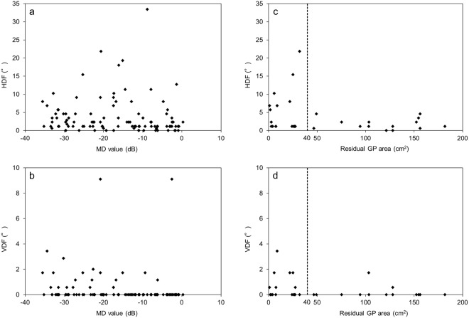
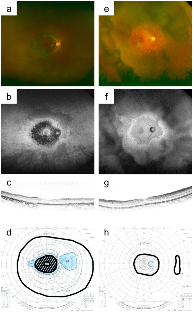
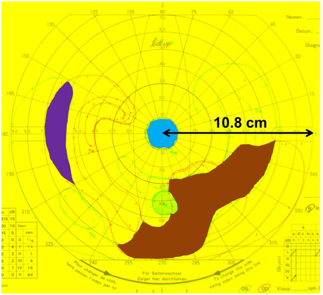
In retinitis pigmentosa (RP), peripheral visual-field loss starts in early stages, whereas central vision loss occurs in advanced stages. Sensory strabismus gradually occurs in RP. We investigated the relationship between ocular deviation and visual function and explored for sensory strabismus risk factors in 119 consecutive patients with RP at various stages. We assessed ocular deviation at far and near distances, that is the central visual field, using the mean deviation (MD) value and visual acuity (VA), and the residual binocular field area, using Goldmann perimetry (GP), in 33 patients. The horizontal ocular deviation at near distance was >10° in 30% patients and correlated with residual visual function. Although there was no effective cut-off value for central visual function, a cut-off residual GP area of 40 cm2 distinguished patients with a larger from those with a smaller horizontal ocular deviation at far distance (P = 0.04). Our findings suggest that visual function is negatively associated with ocular deviation in patients with RP and that the sensory strabismus risk is relatively high for patients with a binocular visual field <40 cm2. Thus, screening for ocular alignment may be necessary for patients with RP-associated severe vision loss as part of their comprehensive care.
Conflict of interest statement
The authors declare no competing interests.
Figures
Similar articles
-
Correlation between macular blood flow and central visual sensitivity in retinitis pigmentosa.Acta Ophthalmol. 2015 Dec;93(8):e644-8. doi: 10.1111/aos.12693. Epub 2015 Feb 17. Acta Ophthalmol. 2015. PMID: 25688697
-
Reduced Central Retinal Artery Blood Flow Is Related to Impaired Central Visual Function in Retinitis Pigmentosa Patients.Curr Eye Res. 2017 Nov;42(11):1503-1510. doi: 10.1080/02713683.2017.1338350. Epub 2017 Sep 14. Curr Eye Res. 2017. PMID: 28910168 Free PMC article.
-
Visual acuity and 10 degrees automated static perimetry in eyes with retinitis pigmentosa.Jpn J Ophthalmol. 2002 Sep-Oct;46(5):581-5. doi: 10.1016/s0021-5155(02)00548-8. Jpn J Ophthalmol. 2002. PMID: 12457920
-
Progression Rate of Visual Function and Affecting Factors at Different Stages of Retinitis Pigmentosa.Biomed Res Int. 2022 Jul 14;2022:7204954. doi: 10.1155/2022/7204954. eCollection 2022. Biomed Res Int. 2022. PMID: 35872870 Free PMC article. Review.
-
Retinitis pigmentosa: visual function and multidisciplinary management.Clin Exp Optom. 2005 Sep;88(5):335-50. doi: 10.1111/j.1444-0938.2005.tb06717.x. Clin Exp Optom. 2005. PMID: 16255692 Review.
Cited by
-
Alteration Ocular Motility in Retinitis Pigmentosa: Case-Control Study.Clin Optom (Auckl). 2024 Feb 22;16:55-69. doi: 10.2147/OPTO.S446717. eCollection 2024. Clin Optom (Auckl). 2024. PMID: 38410094 Free PMC article.
-
Review: Binocular double vision in the presence of visual field loss.J Vis. 2024 Jun 3;24(6):13. doi: 10.1167/jov.24.6.13. J Vis. 2024. PMID: 38899959 Free PMC article. Review.
-
Strabismus in an Adolescent With Stargardt Disease: An Atypical Presentation.Cureus. 2024 Sep 13;16(9):e69325. doi: 10.7759/cureus.69325. eCollection 2024 Sep. Cureus. 2024. PMID: 39398711 Free PMC article.
