. 2018 Oct 5;8(1):14890.
doi: 10.1038/s41598-018-33232-1.

Splenectomy modulates early immuno-inflammatory responses to trauma-hemorrhage and protects mice against secondary sepsis

Affiliations

Splenectomy modulates early immuno-inflammatory responses to trauma-hemorrhage and protects mice against secondary sepsis

S Drechsler et al. Sci Rep. .

Abstract

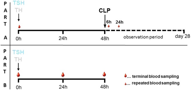
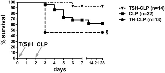
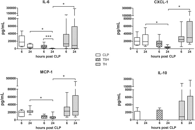
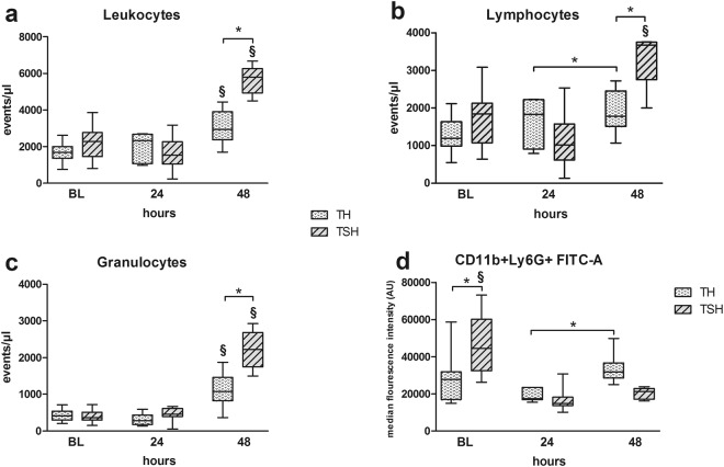
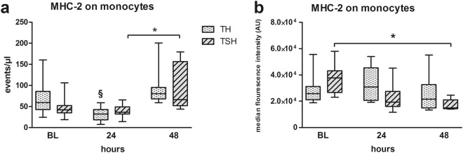
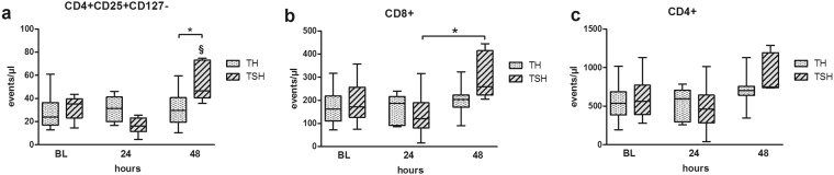
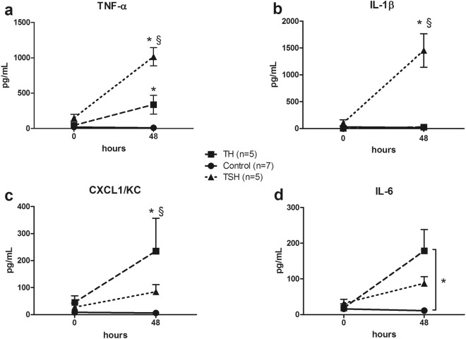
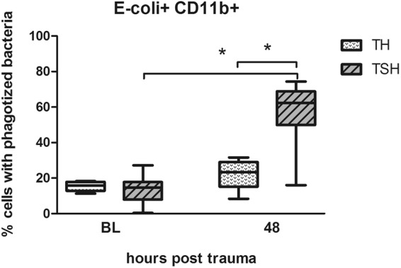
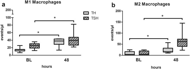
In polytrauma patients, the impact of splenectomy is equivocal, ranging from negative to protective. We investigated the impact of splenectomy on immune responses in the 1st-hit polytrauma alone and on survival in the post-traumatic sepsis (2nd hit). Female BALB/c mice underwent polytrauma (1st hit) consisting of either a) TH: femur fracture, hemorrhagic shock or b) TSH: splenectomy, femur fracture, hemorrhagic shock. Additionally, the polytrauma hit was followed by cecal ligation and puncture (CLP) 48 h later and compared to CLP alone. Splenectomy improved the 28-day survival in secondary sepsis to 92% (from 62%), while TH lowered it to 46% (p < 0.05). The improved survival was concurrent with lower release of inflammatory cytokines (IL-6, CXCL-1, MCP-1) and increase of C5a post-CLP. In the polytrauma hit alone, TSH induced stronger neutrophilia (1.9 fold) and lymphocytosis (1.7 fold) when compared to TH mice. Moreover, TSH resulted in a 41% rise of regulatory T-cells and reduced the median fluorescence intensity of MHC-2 on monocytes by 55% within 48 h (p < 0.05). Conversely, leukocyte phagocytic capacity was significantly increased by 4-fold after TSH despite a similar M1/M2 macrophage profile in both groups. Summarizing, splenectomy provoked both immuno-suppressive and immuno-stimulatory responses but was life-saving in secondary sepsis. Additionally, the polytrauma components in 2-hit models should be tested for their effects on outcome; the presumed end-effect of the 1st hit solely based on the common immuno-inflammatory parameters could be misleading.


Conflict of interest statement

The authors declare no competing interests.

Figures

Figure 1
Figure 1
Experimental Study Design. (Part A):  12-week-old, female BALB/c mice were subjected to either TSH (femur fracture/splenectomy/hemorrhagic shock) or TH (femur fracture/hemorrhagic shock) followed by CLP (cecal ligation and puncture) to induce secondary sepsis 48 h later, or to CLP alone. 20 µl of blood, indicated by small blood drops, were collected at 6 h and 24 h post-CLP and survival was observed for 28 days. (Part B): 12 week old, female BALB/c mice were subjected to either TSH (unilateral femur fracture/splenectomy/hemorrhagic shock) or TH (unilateral femur fracture/hemorrhagic shock). Terminal blood, indicated by large blood drops, was collected either 24 h or 48 h post trauma.
Figure 2
Figure 2
28-day Kaplan Meier Survival Curve. 3 month old, female BALB/c mice were subjected to femur fracture, splenectomy and hemorrhagic shock (TSH, triangle, n = 14) or femur fracture and hemorrhagic shock (TH, circle, n = 13) followed by cecal ligation and puncture (CLP) to induce polymicrobial abdominal sepsis 2 days later, or to CLP alone (square, n = 22). Survival was followed for 28 days. §p < 0.05 compared to all other groups.
Figure 3
Figure 3
Selected plasma cytokines at 6 h and 24 h after TSH/TH-CLP. Female BALB/c mice were subjected to femur fracture, splenectomy and hemorrhagic shock (TSH) or femur fracture and hemorrhagic shock (TH) followed by cecal ligation and puncture (CLP) to induce polymicrobial abdominal sepsis 2 days later, or to CLP alone. Plasma levels of IL-6, IL-10, CXCL-1 and MCP-1 were assessed at 6 h and 24 h after TSH (TSH-CLP) or TH (TH-CLP) followed by secondary CLP or after CLP alone. For CLP: n = 7 per time point, TSH-CLP: n = 9 per time point, TH-CLP: n = 9 per time point. *p < 0.05.
Figure 4
Figure 4
Comparison of leukocyte populations after TSH and TH. Female BALB/c mice were subjected to femur fracture, splenectomy and hemorrhagic shock (TSH) or femur fracture and hemorrhagic shock (TH). (ac) Absolute numbers of circulating leukocytes, lymphocytes and granulocytes at BL (induction of trauma), 24 h and 48 h after TSH or TH. (d) Comparison of median fluorescence activity of activated granulocytes (CD11b+Ly6G+ cells) at BL (induction of trauma), 24 h and 48 h after TSH or TH. For TH: BL n = 24, 24 h n = 16, 48 h n = 12. For TSH: BL n = 11, 24 h n = 6, 48 h n = 5. *p < 0.05, §p < 0.05 compared to all other time-points from the same group (i.e. TSH or TH).
Figure 5
Figure 5
(a) Comparison of MHC-2+ monocytes. Female BALB/c mice were subjected to femur fracture, splenectomy and hemorrhagic shock (TSH) or femur fracture and hemorrhagic shock (TH). (a) MHC-2 expressing monocytes at BL (induction of trauma), 24 h and 48 h after TSH and TH. B. Median fluorescence intensity of MHC-2 expression on monocytes at BL (induction of trauma), 24 h and 48 h after TSH or TH. For TH: BL n = 7, 24 h n = 16, 48 h n = 12. For TSH: BL n = 11, 24 h n = 6, 48 h n = 5, *p < 0.05, §p < 0.05 compared to all other time-points from the same group (i.e. TSH or TH).
Figure 6
Figure 6
Comparison of T-cell populations. Female BALB/c mice were subjected to femur fracture, splenectomy and hemorrhagic shock (TSH) or femur fracture and hemorrhagic shock (TH). Absolute numbers of circulating (a). CD4+CD25+CD127− T-cells, (b). CD8+ T-cells and (c). CD4+ T-cells at BL (induction of trauma), 24 h and 48 h time point after TSH or TH. For TH: at BL n = 24, 24 h n = 16, 48 h n = 12. For TSH: BL n = 11, 24 h n = 6, 48 h n = 5. *p < 0.05.
Figure 7
Figure 7
Ex-vivo cytokine release upon LPS stimulation of the whole blood. Female BALB/c mice were subjected to femur fracture, splenectomy and hemorrhagic shock (TSH) or femur fracture and hemorrhagic shock (TH) and whole blood was collected at BL and 48 h. (ad) TNFα, IL-1β, IL-6 and CXCL-1 release upon whole blood stimulation with 10 µg LPS at 48 h after TH (square) or TSH (triangle) compared to healthy control group. (ac) *p < 0.05 between trauma models, §p < 0.05 compared to both groups. (d) *p < 0.05 Data presented as mean ± SD.
Figure 8
Figure 8
Complement Component C5a. Female BALB/c mice were subjected to femur fracture, splenectomy and hemorrhagic shock (TSH) or femur fracture and hemorrhagic shock (TH) and whole blood was collected at BL, 24 and 48 h to assess C5a levels from 1:10 diluted plasma samples using ELISA. For TSH at BL n = 12, at 24 h n = 12, at 48 h n = 5, for TH n = 11 for all time points. *p < 0.05.
Figure 9
Figure 9
Phagocytosis assay. Female BALB/c mice were subjected to femur fracture, splenectomy and hemorrhagic shock (TSH) or femur fracture and hemorrhagic shock (TH) and whole blood was collected at BL and 48 h. Phagocytic uptake of pHrodo® labeled E. coli by CD11b+ cells was detected at baseline and 48 h after TSH and TH when incubated for 24 h at 0 °C. For TSH at BL n = 19, at 48 h n = 8, for TH n = 8 for both time points. *p < 0.05.
Figure 10
Figure 10
Macrophage polarization. Female BALB/c mice were subjected to femur fracture, splenectomy and hemorrhagic shock (TSH) or femur fracture and hemorrhagic shock (TH) and whole blood was collected at BL and 48 h to investigate changes in the macrophage polarization. (a) M1 macrophages, (b) M2 Macrophages. For TSH n = 8 and for TH n = 7 for both time points. *p < 0.05.

References

    1. Marik PE, Flemmer M. The immune response to surgery and trauma: Implications for treatment. J Trauma Acute Care Surg. 2012;73:801–808. doi: 10.1097/TA.0b013e318265cf87. - DOI - PubMed
    1. Lenz A, Franklin GA, Cheadle WG. Systemic inflammation after trauma. Injury. 2007;38:1336–1345. doi: 10.1016/j.injury.2007.10.003. - DOI - PubMed
    1. Manson J, Thiemermann C, Brohi K. Trauma alarmins as activators of damage-induced inflammation. Br J Surg. 2012;99(Suppl 1):12–20. doi: 10.1002/bjs.7717. - DOI - PubMed
    1. Brøchner Anne, Toft Palle. Pathophysiology of the systemic inflammatory response after major accidental trauma. Scandinavian Journal of Trauma, Resuscitation and Emergency Medicine. 2009;17(1):43. doi: 10.1186/1757-7241-17-43. - DOI - PMC - PubMed
    1. Vanzant EL, et al. Persistent inflammation, immunosuppression, and catabolism syndrome after severe blunt trauma. J Trauma Acute Care Surg. 2014;76:21–29. doi: 10.1097/TA.0b013e3182ab1ab5. - DOI - PMC - PubMed

Publication types