. 2019 Mar 1;527(4):767-779.
doi: 10.1002/cne.24552. Epub 2018 Nov 14.

Seasonal changes in neuronal turnover in a forebrain nucleus in adult songbirds

Affiliations

Seasonal changes in neuronal turnover in a forebrain nucleus in adult songbirds

Tracy A Larson et al. J Comp Neurol. .

Abstract

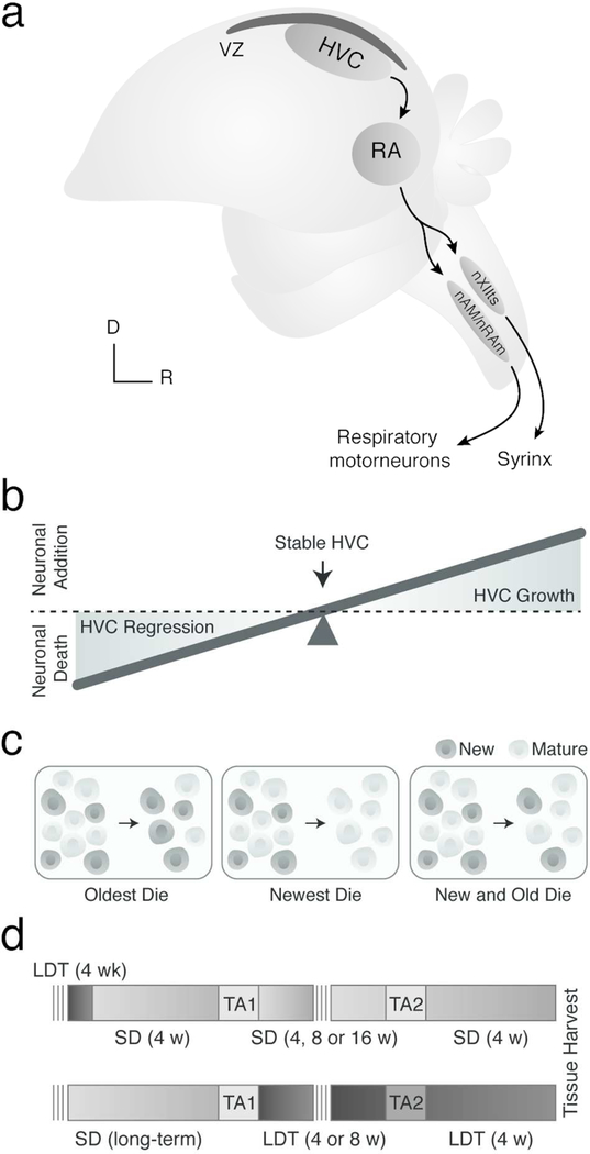
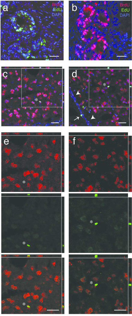
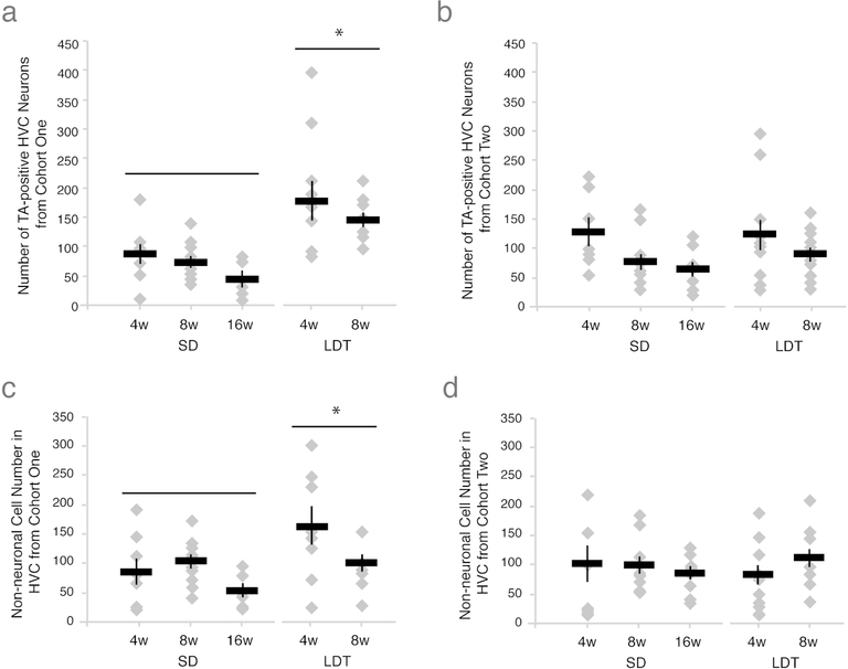
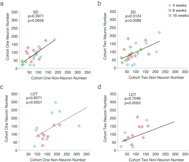
Neuronal death and replacement, or neuronal turnover, in the adult brain are one of many fundamental processes of neural plasticity. The adult avian song control circuit provides an excellent model for exploring mature neuronal death and replacement by new neurons. In the song control nucleus, HVC of adult male Gambel's white-crowned sparrows (Zonotrichia leucophrys gambelli) nearly 68,000 neurons are added each breeding season and die during the subsequent nonbreeding season. To accommodate large seasonal differences in HVC neuron number, the balance between neuronal addition and death in HVC must differ between seasons. To determine whether maintenance of new HVC neurons changes within and between breeding and nonbreeding conditions, we pulse-labeled two different cohorts of new HVC neurons under both conditions and quantified their maintenance. We show that the maintenance of new HVC neurons, as well as new nonneuronal cells, was higher at the onset of breeding conditions than at the onset of nonbreeding conditions. Once a steady-state HVC volume and neuronal number were attained in either breeding or nonbreeding conditions, neuronal and nonneuronal maintenance were similarly low. We found that new neuronal number correlated with a new nonneuronal number within each cohort of new neurons. Together, these data suggest that sex steroids promote the survival of an initial population of new neurons and nonneuronal cells entering HVC. However, once HVC is fully grown or regressed, neuronal and nonneuronal cell turnover is regulated by a common mechanism likely independent of direct sex steroid signaling.

Keywords: adult neurogenesis; anti-BrdU (RRID: AB_2536432); anti-NeuN (RRID: AB_2532109); degeneration; neuronal death; seasonal plasticity; songbird; steroid hormone; testosterone.


Conflict of interest statement

Conflict of Interest: The authors declare no competing financial interests.

Figures

Figure 1.
Figure 1.
(A) Schematic illustrating the major projections within the song production circuit. Note the ventricular zone (VZ) in black directly dorsal to HVC. RA = robust nucleus of the arcopallium, nXII = tracheosyringeal portion of hypoglossal nucleus, nAM/nRAm = nucleus ambiguus and nucleus retroambigualis, D = dorsal, and R = rostral. HVC grows and regresses each breeding and nonbreeding season by about 68,000 neurons as a result of increased new neuronal survival and neuronal apoptosis, respectively. (B) Schematic illustrating how the balance between neuronal birth and death hypothetically contributes to HVC growth and regression. (C) Predictive models for HVC neuronal turnover. New neurons that enter HVC might: entirely replace oder neurons (left), temporarily contribute to HVC growth and then die (middle), or serve to replace some older neurons, but also die (right). (D) Experimental design for testing the predictive models above in nonbreeding (i.e., top) and breeding (bottom) conditions. SD = short day photoperiods to promote nonbreeding physiological condition (light grey), LDT = long day photoperiods and exogenous testosterone pellet to stimulate breeding-like physiological condition (dark grey), TA1 and TA2 = thymidine analog 1 and thymidine analog 2. Note: darker grey TA2 background (bottom) indicates injections were administered in LDT conditions rather than SD conditions.
Figure 2.
Figure 2.
Photomicrographs of thymidine labeled cells. (A) Image of intestinal tissue labeled for EdU (green), BrdU (red), and DAPI (blue) from bird injected with EdU two hours prior to tissue harvest. The BrdU antibody did not cross react with EdU positive cells, confirming that the MoBU BrdU antibody against BrdU does not react with EdU labeled DNA. (B) Image of intestine labeled for EdU (green), BrdU (red), and DAPI, a nuclear marker (blue) from bird injected with BrdU two hours prior to tissue harvest. The MoBU antibody labels BrdU positive cells, whereas the EdU click reaction does not. (C) Representative image of a thymidine analog positive neuron in HVC. Thymidine analog label, in this case, EdU, is shown in green, while label for NeuN, a neuronal marker, is shown in red, and DAPI, a nuclear marker, is shown in blue. The white star indicates a new neuron co-labeled with EdU, NeuN, and DAPI in the nucleus, as also observed in the cut outs of the X-Z and Y-Z planes. The grey box indicates the area of zoom in (E). (D) Representative image of thymidine analog positive non-neuronal cells in the proliferative ventricular zone (VZ) and HVC. Thymidine analog label, in this case, BrdU, is shown in green, NeuN in red, and DAPI in blue. The white arrows heads indicate the VZ, whereas the white arrow indicates a BrdU-positive cell that retained label (a very slowly dividing NSC or a postmitotic non-neuronal cell) within the VZ. The white star indicates a BrdU-positive non-neuronal cells within HVC. The box highlights the region of zoom in (F). (E) Magnified region from (B) showing an EdU-positive neuron in HVC. Single channel images for NeuN positive cells (top, red), the EdU positive cell (middle, green), and co-label (bottom). The presence of NeuN label both within and immediately outside of the area labeled with EdU (see X-Z and Y-Z cutouts) confirm neuronal identity of this new cell. (F) Magnified region from (C) showing an BrdU-positive non-neuronal cell in HVC. Single channel images for NeuN positive cells (top, red), the BrdU positive cell (middle, green), and co-label (bottom). The absence of NeuN label within the area labeled with BrdU (see X-Z and Y-Z cutouts) suggests that this BrdU labeled cell is nonneuronal. All scale bars (white) are 20 μm.
Figure 3.
Figure 3.
Breeding conditions promote maintenance of an initial cohort of new neurons and non-neuronal cells, but do not alter subsequent neuronal turnover. (A) Number of thymidine analog positive neurons in one hemisphere of HVC from cohort one across all survival times in nonbreeding and breeding condition. Depicted results include both BrdU and EdU positive cell data pooled within experimental group (see Table 2 for lack of thymidine analog order effect), with data from individual birds represented as grey diamonds and the group mean as a black bar with S.E.M.. The number of new HVC neurons maintained from the initial cohort of thymidine analog label are significantly higher under breeding conditions, suggesting that LDT conditions promote greater maintenance of an initial cohort of new neurons than in SD. Because in both nonbreeding and breeding conditions neurons from the first cohort are maintained, these data further indicate that some new neurons replace older neurons, while other new neurons die (see Figure 1C, right panel). (B) Number of new neurons maintained in HVC on one side of the brain from the second thymidine analog label in all experimental groups. Physiological condition and interval between injection of the two thymidine analogs did not significantly affect the number of new neurons from the second cohort maintained in HVC. These data suggest that during stable conditions, new neurons enter HVC to replace older neurons in equal amounts regardless of condition (Tables 2 and 3). (C-D) The number of new non-neuronal HVC cells maintained from the cohort labeled with the first thymidine analog (C) and the second thymidine analog (D) across all survival times in nonbreeding and breeding condition. LDT conditions lead to greater maintenance of new non-neuronal cells from the first thymidine analog-labeled cohort but not the second when compared to SD conditions (Table 3). These data suggest that breeding conditions not only promote the increased maintenance of an initial population of new neurons but also new non-neuronal cells, which together contribute to the growth of HVC.
Figure 4.
Figure 4.
The number of new neuronal and non-neuronal cells co-vary significantly within, but not across, cohorts (also see Table 4). The results of Spearman’s rank correlation from comparisons of: (A) neuronal and non-neuronal cells labeled with the first thymidine analog in SD, (B) neuronal and non-neuronal cells labeled with the second TA in SD, (C) neuronal and non-neuronal cells labeled with the first thymidine analog in LDT, (D) neuronal and non-neuronal cells labeled with the second thymidine analog in LDT. The correlation between new neuronal and non-neuronal cells within, but not across, thymidine analog-labeled cohorts suggests that a temporally controlled common factor present during both breeding and nonbreeding conditions mediates addition, survival, or both of new cell populations.

References

    1. Alvarez-Buylla A, Ling CY, Yu WS. 1994. Contribution of neurons born during embryonic, juvenile, and adult life to the brain of adult canaries: regional specificity and delayed birth of neurons in the song-control nuclei. J Comp Neurol 347(2):233–248. - PubMed
    1. Alvarez-Buylla A, Theelen M, Nottebohm F. 1990. Proliferation “hot spots” in adult avian ventricular zone reveal radial cell division. Neuron 5(1):101–109. - PubMed
    1. Barnea A, Pravosudov V. 2011. Birds as a model to study adult neurogenesis: bridging evolutionary, comparative and neuroethological approaches. Eur J Neurosci 34(6):884–907. - PMC - PubMed
    1. Bentley GE, Audage NC, Hanspal EK, Ball GF, Hahn TP. 2003. Photoperiodic response of the hypothalamo-pituitary-gonad axis in male and female canaries, Serinus canaria. J Exp Zool A Comp Exp Biol 296(2):143–151. - PubMed
    1. Brenowitz EA. 2008. Plasticity of the song control system in adult birds In: Zeigler HP, Marler P, eds. Neuroscience of birdsong. Cambridge: Cambridge University Press; p 332–349.

Publication types