. 2018 Nov 26;57(48):15675-15680.
doi: 10.1002/anie.201806901. Epub 2018 Oct 30.

A Single Extracellular Vesicle (EV) Flow Cytometry Approach to Reveal EV Heterogeneity

Affiliations

A Single Extracellular Vesicle (EV) Flow Cytometry Approach to Reveal EV Heterogeneity

Wen Shen et al. Angew Chem Int Ed Engl. .

Abstract

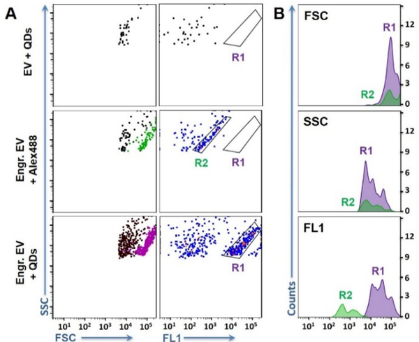
Extracellular vesicles (EVs) actively participate in intercellular communication and pathological processes. Studying the molecular signatures of EVs is key to reveal their biological functions and clinical values, which, however, is greatly hindered by their sub-100 nm dimensions, the low quantities of biomolecules each EV carries, and the large population heterogeneity. Now, single-EV flow cytometry analysis is introduced to realize single EV counting and phenotyping in a conventional flow cytometer for the first time, enabled by target-initiated engineering (TIE) of DNA nanostructures on each EV. By illuminating multiple markers on single EVs, statistically significant differences are revealed among the molecular signatures of EVs originating from several breast cancer cell lines, and the cancer cell-derived EVs among the heterogeneous EV populations are successfully recognized. Thus, our approach holds great potential for various biological and biomedical applications.

Keywords: engineering; flow cytometry analysis; heterogeneity; molecular signatures; single extracellular vesicle analysis.


Figures

Figure 1.
Figure 1.
A) Schematic of the Single Extracellular Vesicle Flow Cytometry Analysis technique enabled by target-initiated engineering of DNA nanostructures. B) Analysis of the long DNA products by gel electrophoresis: Lane 1 - reaction probes only; Lane 2, 3, and 4 - reaction triggered by the simple initiator, CD63 protein, and CD63+ EVs, respectively. C) TEM image of the Engineered EV (Engr. EV). D) Fluorescence microscopy image of the Engr. EV tagged with rhodamine-nanoparticles (Engr. EV-RhNPs; shown in red) or QDs-525 (Engr. EV-QDs shown in green). Circles – Engr. EV-RhNPs or Engr. EV-QDs, well separated from each other; Square – one Engr. EV-QDs located nearby another Engr. EV-RhNPs; Triangle – Engr.EV labelled with both QDs and Rh-NPs.
Figure 2.
Figure 2.
A) Representative scatter plots of flow cytometry analysis of the EVs before and after TIE. Top to bottom are: Standard EVs directly labelled with QDs (EV + QDs); Engineered EVs labelled with Alexa488 or QDs-525. The particle populations shown in green and purple on the light scatter plots of FSC vs. SSC were those included in R2 and R1, respectively, on the flow plots of FL1 vs. SSC. B) Histograms for the signals of FSC, SSC, and fluorescence produced by the engineered EVs labelled with Alexa488 (green) and QDs-525 (purple). All samples started with ~ 109 EV particles.
Figure 3.
Figure 3.
A) Representative flow cytometry plots of the particle cluster determined by the relative expression levels of HER2 and CD63 for the EVs from different cell lines. B) Scheme of the dual hybridization cascade system for recognition of two markers on the same EV. C) Box chart of the mean fluorescence intensity (MFI) ratio between FL4 and FL1 of the EVs from three cell lines. *p < 0.05, and **p < 0.01, n = 5. IQR - interquartile range. D) Flow cytometry scatter plots and fluorescence histograms for analysis of EV mixtures. R1 and R3 are defined in Fig. 3A using the EVs from the corresponding cell lines. The particle clusters were coloured on the scatter plot of FSC vs. SSC and the histograms based on the gates defined in the HER2 vs. CD63 plots.

References

    1. Antonyak MA, Cerione RA, Proc. Natl. Acad. Sci. USA 2015, 112, 3589–3590. - PMC - PubMed
    2. Brown L, Wolf JM, Prados-Rosales R, Casadevall A, Nat. Rev. Microbiol. 2015, 13, 620–630. - PMC - PubMed
    1. Sullivan LB, Nat. Chem. Bio. 2017, 13, 924–925. - PubMed
    2. Thompson AG, Gray E, Heman-Ackah SM, Mäger I, Talbot K, Andaloussi SE, Wood MJ, Turner MR, Nat. Rev. Neurol. 2016, 12, 346–357. - PubMed
    1. Ruivo CF, Adem B, Silva M, Melo SA, Cancer Res. 2017, 77, 6480–6488. - PubMed
    2. Han L, Xu J, Xu Q, Zhang B, Lam EW, Sun Y, Med. Res. Rev. 2017, 37, 1318–1349. - PubMed
    1. Tkach M, Thery C, Cell 2016, 164, 1226–1232. - PubMed
    1. Jiang Y, Shi M, Liu Y, Wan S, Cui C, Zhang L, Tan W, Angew. Chem. Int. Ed. 2017, 56, 11916–11920. - PMC - PubMed
    2. Greening DW, Xu R, Ji H, Tauro BJ, Simpson RJ, Methods Mol. Biol. 2015, 1295, 179–209. - PubMed
    3. Pocsfalvi G, Stanly C, Fiume I, Vekey K, Chromatogr JA 2016, 1439, 26–41. - PubMed
    4. Zhao Z, Yang Y, Zeng Y, He M, Lab Chip 2016, 16, 489–496. - PMC - PubMed
    5. Rider MA, Hurwitz SN, Meckes DG, Sci. Rep. 2016, 6, 23978. - PMC - PubMed

Publication types