. 2019 Mar;21(3):e12961.
doi: 10.1111/cmi.12961. Epub 2018 Nov 20.

Role of clathrin-mediated endocytosis in the use of heme and hemoglobin by the fungal pathogen Cryptococcus neoformans

Affiliations

Role of clathrin-mediated endocytosis in the use of heme and hemoglobin by the fungal pathogen Cryptococcus neoformans

Gaurav Bairwa et al. Cell Microbiol. 2019 Mar.

Abstract

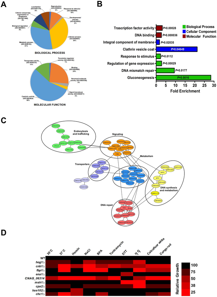
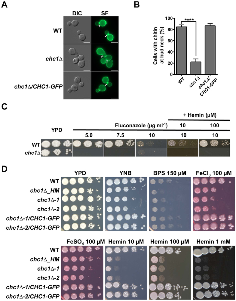
Heme is a major source of iron for pathogens of humans, and its use is critical in determining the outcome of infection and disease. Cryptococcus neoformans is an encapsulated fungal pathogen that causes life-threatening infections in immunocompromised individuals. C. neoformans effectively uses heme as an iron source, but the underlying mechanisms are poorly defined. Non-iron metalloporphyrins (MPPs) are toxic analogues of heme and are thought to enter microbial cells via endogenous heme acquisition systems. We therefore carried out a mutant screen for susceptibility against manganese MPP (MnMPP) to identify new components for heme uptake in C. neoformans. We identified several genes involved in signalling, DNA repair, sugar metabolism, and trafficking that play important roles in susceptibility to MnMPP and in the use of heme as an iron source. We focused on investigating the role of clathrin-mediated endocytosis (CME) and found that several components of CME including Chc1, Las17, Rvs161, and Rvs167 are required for growth on heme and hemoglobin and for endocytosis and intracellular trafficking of these molecules. We show that the hemoglobin uptake process in C. neoformans involves clathrin heavy chain, Chc1, which appears to colocalise with hemoglobin-containing vesicles and to potentially assist in proper delivery of hemoglobin to the vacuole. Additionally, C. neoformans strains lacking Chc1, Las17, Rvs161, or Rvs167 were defective in the elaboration of several key virulence factors, and a las17 mutant was avirulent in a mouse model of cryptococcosis. Overall, this study unveils crucial functions of CME in the use of heme iron by C. neoformans and reveals a role for CME in fungal pathogenesis.

Keywords: CHC1; Cryptococcus; clathrin; endocytosis; heme; hemoglobin; iron.


Figures

Figure 1:
Figure 1:. Identification of cryptococcal mutants with altered susceptibility to manganese metalloporphyrin (MnMPP).
(A) Growth profile at 72 h for iron-starved or non-starved WT cells in YNB medium containing different concentrations of either manganese or gallium metalloporphyrin. The results are the means ± standard errors of the mean (SEM) from three independent experiments. (***P < 0.001). (B) Graphical outline of the strategy used to screen the deletion mutant libraries against MnMPP. Plate A; YNB, Plate B; YNB with BPS (150 μM), Plate C; YNB with BPS (150 μM) and hemin (10 μM) [hemin media] and Plate D; YNB with BPS (150 μM), hemin (10 μM) and MnMPP (10 μM) [MnMPP media]. (C) Growth profile of individual strains of the three libraries screened against MnMPP, representing the relative growth differences in percentage between the MnMPP and hemin media conditions at 72 h. The blue line represents the average relative growth (ARG) value calculated from the relative growth difference values of all the strains in a library. The red line marks the value which is 50% of the ARG, while the green line marks the value at 150% of the ARG. Circles filled with green color highlight some of the interesting mutants showing resistance to MnMPP. Red filled circles represent some of the interesting mutants that are sensitive to MnMPP.
Figure 2:
Figure 2:. Gene Ontology and interaction analyses of genes involved in MnMPP resistance.
(A) Gene Ontology analysis of the genes involved in MnMPP resistance for the categories of biological process and molecular functions. The H99 genes were matched to the C. neoformans JEC21 gene IDs retrieved from the FungiDB (http://fungidb.org/fungidb/) database and analyzed with the PANTHER gene ontology online tool (http://pantherdb.org) using C. neoformans as the organism. (B) Functional enrichment analysis of genes involved in MnMPP resistance. Genes were analyzed for enrichment of the GO biological process, molecular function and cellular component categories. Enriched groups were scored by comparison to a background list of the C. neoformans H99 genome database using a cut-off of P < 0.05 and Bonferroni correction. (C) STRING network analysis of the C. neoformans genes involved in MnMPP resistance. Interaction network details were obtained with JEC21 gene IDs from the STRING online tool (https://string-db.org). The network was extracted and visualized with Cytoscape 3.6.1 software (http://www.cytoscape.org) and clustering was performed with the clusterMaper plugin using the community cluster (GLay) network algorithm. Individual clusters are marked with an elliptical circle. (D) Heatmap depicting the growth behavior of selected MnMPP resistant mutants in various environmental stress conditions. Growth of the individual strains at 30°C, 37°C and in presence of hemin (10 μM), NaCl (1.5 M), brefeldin A; BFA (20 μg ml−1), tunicamycin (0.15 μg ml−1), dithiothreitol; DTT (10 mM), H2O2 (1 mM), calcofluor white (1 mg ml−1), and congo red (2.5 mg ml−1) was checked and displayed with color gradients (indicated as percentages of the WT).
Figure 3:
Figure 3:. Sequence analysis of the clathrin heavy chain 1 protein in fungi and higher eukaryotes
(A) Predicted domain architecture of the C. neoformans Chc1 protein and multiple alignment of the 1st Clathrin Heavy Chain Repeat (CHCR) sequences of the Chc1 proteins from selected organisms. The amino acid sequence alignment was performed with Clustal W (http://www.ch.embnet.org); identical residues are boxed in black (indicated with *) and similar residues are shown in gray (indicated with ●). The numbers on the left represent the starting position of the CHCR repeat sequence in the protein from the respective organism. (B) Phylogenetic tree of the Chc1 proteins from selected organisms. The phylogenetic analysis of the Chc1 protein was performed in MEGA7 (https://www.megasoftware.net) by using the Maximum Likelihood method and applying Neighbor-Join and BioNJ algorithms. The tree is drawn to scale, with branch lengths measured in the number of substitution per site. CnA, C. neoformans var. grubii, H99 (serotype A); CnD, C. neoformans var. neoformans JEC21 (serotype D); Cg, C. gattii; Sc, Saccharomyces cerevisiae; Ca, Candida albicans; Hm, Homo sapiens; Mm, Mus musculus.
Figure 4:
Figure 4:. A C. neoformans chc1∆ mutant exhibits altered cell morphology and an inability to grow in presence of fluconazole and heme-iron sources.
(A) Representative differential interference contrast (DIC) and fluorescent images of WT, chc1∆ and chc1∆/CHC1-GFP complemented cells stained with Solophenyl Flavine (SF) to visualize the cell architecture and chitin at the cell wall. White arrows indicate the presence or absence of chitin deposition at the mother-daughter bud neck region in the indicated strains. Scale bar = 5 μm. (B) Quantification of the cell population having deposition of chitin at the mother-daughter bud neck region in the indicated strains. A total of 100 cells for each strain were counted. Data represent the mean ± standard deviation (SD) of two independent experiments. (****P < 0.0001). (C) and (D) Spot assays of 10-fold serial dilutions of the indicated strains on medium supplemented as shown. Cells of the indicated strains were starved for iron for 24 h prior to the spotting. All the plates were incubated for 3-4 days at 30°C before being photographed. The mutant designated chc1∆_HM is from the UCSF-2015 deletion collection and the other two chc1∆ mutants were constructed as described in the Experimental Procedures. YNB; Yeast Nitrogen Base, BPS; Bathophenanthroline disulfonate.
Figure 5:
Figure 5:. The C. neoformans chc1∆ mutant cannot use hemin and hemoglobin as sole iron sources.
Cells of the indicated strains were starved for iron for 24 h and then transferred to YNB-LIM medium containing either hemin or hemoglobin. Growth at 30°C was monitored by recording OD600 over a 96-h time course at the indicated time intervals. Data are represented as the mean ± SEM of 3-4 independent experiments.
Figure 6:
Figure 6:. The C. neoformans Chc1 protein is required for endocytosis and intracellular trafficking.
(A) C. neoformans Chc1 was tagged with GFP at its C-terminus and its localization was visualized in exponentially growing cells under a fluorescence microscope using the EGFP filter. White arrowheads indicate the Chc1-GFP puncta. Images are the representative of two independent experiments. Scale bar = 2 μm. (B) The absence of CHC1 results in a significant delay in FM4-64 endocytosis and trafficking to the vacuole. YPD-grown cells of the indicated strains were labelled with FM4-64 on ice for 20 min and were visualized under a fluorescence microscope after the indicated times of incubation at room temperature. FM4-64 was visualized using a rhodamine filter. Images are representative of two independent experiments. Scale bar = 5 μm. (C) Chc1 in C. neoformans colocalizes with FM4-64-labelled structures. The WT strain expressing Chc1-GFP was labelled with FM4-64 and visualized under a fluorescence microscope after the indicated times of incubation at room temperature. Images are the representative of two independent experiments. Scale bar = 2 μm.
Figure 7:
Figure 7:. Chc1 is required for the endocytosis and trafficking of hemoglobin in C. neoformans.
(A) To assess the uptake of labelled hemoglobin (HbAlexa594), WT, chc1∆ and chc1∆/CHC1-GFP complemented strains were incubated with HbAlexa594 and the cell-wall labeling dye Fluorescent Brightener 28 (FB-28) at 4°C and subsequently shifted to room temperature for the indicated times. Representative images of three independent experiments are shown. White triangles indicate the presence of HbAlexa594. Scale bar = 5 μm. (B) Chc1 in C. neoformans colocalizes with hemoglobin containing vesicles. The WT strain expressing Chc1-GFP was incubated with HbAlexa594 and visualized under a fluorescence microscope at the indicated time points of incubation at room temperature. Representative images of three independent experiments are shown. The white triangles indicate the colocalization of Chc1-GFP and HbAlexa594. Scale bar = 2 μm. (C) Quantification of the Chc1-GFP expressing cell population showing HbAlexa594 labeling (HbAlexa594 +ve) and colocalization of Chc1-GFP and HbAlexa594 (HbAlexa594 +ve and GFP +ve) after 10 and 30 min. Data represent the analysis for at least 50 cells with mean ± SEM of three independent experiments. (**P < 0.01). (D) The chc1∆ strain exhibits reduced intracellular heme levels. Total heme content was measured from the cells grown overnight in rich YPD medium and is represented relative to the WT levels. C. neoformans atm1∆ and atm1∆/ATM1-GFP complemented strains were used as the controls. Data represent the mean ± SEM of 4 independent experiments.
Figure 8:
Figure 8:. The C. neoformans chc1∆ mutant exhibits diminished capsule and melanin production and is sensitive to several stress conditions.
(A) DIC microscopy images of India ink-stained cells grown in capsule-induction medium as described in the Experimental Procedures. Scale bar = 5 μm. (B) Quantification of cell and capsule size following incubation in capsule-induction medium. Data represent the analysis for at least 60 cells for each strain with circles indicating individual data points and the black bar indicating the mean. (***P < 0.001). (C) 10-fold serial dilution assay of the indicated strains on L-DOPA agar plates to assess melanin production. Plates were incubated for 3 days at 30°C before being photographed. (D) The indicated strains were grown in liquid L-DOPA medium at 30°C for 24 h and melanin secretion was quantified by measuring the OD405 in the supernatant fraction. Data represent the mean ± SEM of three independent experiments. (***P < 0.001) (E) 10-fold serial dilution assay of the indicated strains on medium containing BFA (20 μg ml−1), monensin (1.25 mg ml−1), tunicamycin (0.15 μg ml−1), glycerol (2 %), ethanol (2 %), sodium acetate (1 or 2 %), H2O2 (1.5 mM), LiCl (100 mM), NaCl (1.5 M), KCl (1.5 M), congo red (2.5 mg ml−1) and calcofluor white (1 mg ml−1). All the plates were incubated for 3-4 days at 30°C or 37°C before being photographed.
Figure 9:
Figure 9:. Las17, Rvs161 and Rvs167 are also required for heme-iron utilization in C. neoformans.
(A) 10-fold serial dilutions of iron-starved cells of the indicated strains were spotted on medium supplemented as shown including BPS (100 μM), FeCl3 (100 μM), FeSO4 (100 μM) and hemin (100 or 500 μM). All of the plates were incubated for 3-4 days at 30°C before being photographed. (B) Iron-starved cells of the indicated strains were transferred to YNB-LIM medium containing either hemin or hemoglobin and their growth at 30°C was monitored by recording OD600 over a 96-h time-course at the indicated time intervals. Data are presented as the mean ± SEM of 3-4 independent experiments.
Figure 10:
Figure 10:. Clathrin-mediated endocytosis component Las17 is required for capsule and melanin production, and for virulence.
(A) 10-fold serial dilutions of the strains were spotted on the indicated medium supplemented with different compounds as described in the legend of Figure 8E, with sodium acetate used at a concentration of 2%. All the plates were incubated for 3-4 days at 30°C or 37°C before being photographed. (B) Quantification of C. neoformans cell and capsule size following incubation in capsule-induction medium. Data represent the analysis of at least 60 cells for each strain with circles indicating individual data points and the black bar indicating the mean. (***P < 0.001). (C) 10-fold serial dilutions assay of the indicated strains on L-DOPA agar plates to assess the melanin production. Plates were incubated for 3 days at 30°C or 37°C before being photographed. (D) 10 female BALB/c mice were inoculated intranasally with 2 × 10⁵ cells of each of the strains indicated, and the survival of the mice was monitored daily. Survival differences between groups of mice were evaluated by log-rank tests. The p-values for the mice infected with the WT and mutant strains were statistically different (p < 0.001). Note that the experiment was terminated at 50 days when the mice infected with the mutants became ill.

References

    1. Agarwal S, Rastogi R, Gupta D, Patel N, Raje M, and Mukhopadhyay A (2013) Clathrin-mediated hemoglobin endocytosis is essential for survival of Leishmania. Biochim Biophys Acta BBA - Mol Cell Res 1833: 1065–1077. - PubMed
    1. Almeida F, Wolf JM, and Casadevall A (2015) Virulence-Associated Enzymes of Cryptococcus neoformans. Eukaryot Cell 14: 1173–1185. - PMC - PubMed
    1. Baek Y-U, Li M, and Davis DA (2008) Candida albicans Ferric Reductases Are Differentially Regulated in Response to Distinct Forms of Iron Limitation by the Rim101 and CBF Transcription Factors. Eukaryot Cell 7: 1168–1179. - PMC - PubMed
    1. Baggett JJ, and Wendland B (2001) Clathrin Function in Yeast Endocytosis: Clathrin Function in Yeast Endocytosis. Traffic 2: 297–302. - PubMed
    1. Bahn Y-S, Kojima K, Cox GM, and Heitman J (2005) Specialization of the HOG Pathway and Its Impact on Differentiation and Virulence of Cryptococcus neoformans. Mol Biol Cell 16: 2285–2300. - PMC - PubMed

Publication types

LinkOut - more resources