

Endothelins in inflammatory neurological diseases
- PMID: 30291906
- PMCID: PMC6348026
- DOI: 10.1016/j.pharmthera.2018.10.001
Endothelins in inflammatory neurological diseases
Abstract
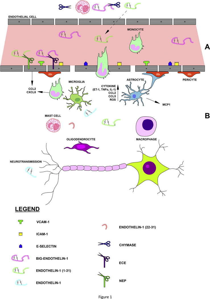
Endothelins were discovered more than thirty years ago as potent vasoactive compounds. Beyond their well-documented cardiovascular properties, however, the contributions of the endothelin pathway have been demonstrated in several neuroinflammatory processes and the peptides have been reported as clinically relevant biomarkers in neurodegenerative diseases. Several studies report that endothelin-1 significantly contributes to the progression of neuroinflammatory processes, particularly during infections in the central nervous system (CNS), and is associated with a loss of endothelial integrity at the blood brain barrier level. Because of the paucity of clinical trials with endothelin-1 antagonists in several infectious and non-infectious neuroinflammatory diseases, it remains an open question whether the 21 amino acid peptide is a mediator/modulator rather than a biomarker of the progression of neurodegeneration. This review focuses on the potential roles of endothelins in the pathology of neuroinflammatory processes, including infectious diseases of viral, bacterial or parasitic origin in which the synthesis of endothelins or its pharmacology have been investigated from the cell to the bedside in several cases, as well as in non-infectious inflammatory processes such as neurodegenerative disorders like Alzheimers Disease or central nervous system vasculitis.
Keywords: Blood-brain barrier (BBB); Central nervous system; Cerebral blood flow (CBF); Chymase; Cytokines; Endothelin subtype A receptor (ET(A)); Endothelin subtype B receptor (ET(B)); Endothelin-1 (ET-1); Mast cells.
Copyright © 2018 Elsevier Inc. All rights reserved.
Conflict of interest statement
Conflict of Interest Statement
The authors declare that there are no conflicts of interest
Figures
References
-
- Adams CW, Poston RN, Buk SJ, Sidhu YS, & Vipond H (1985). Inflammatory vasculitis in multiple sclerosis. J Neurol Sci, 69, 269–283. - PubMed
-
- Adams S, Brown H, & Turner G (2002). Breaking down the blood-brain barrier: signaling a path to cerebral malaria? Trends Parasitol, 18, 360–366. - PubMed
-
- Alencar A, & Elejalde P (1960). O sistema nervoso central na infestaçao experimental do camundongo albino pelo Schizotrypanum cruzi. J Brasil Neurol, 12, 49–57.
-
- Alencar A (1964). [Clinical and Biological Aspects of Neurological and Muscular Manifestations of South American Trypanosomiasis (Chagas’ Disease)]. Rev Neuropsiquiatr, 27, 57–69. - PubMed
Publication types
MeSH terms
Substances
Grants and funding
LinkOut - more resources
Full Text Sources
Medical
