. 2018 Oct 6;17(1):146.
doi: 10.1186/s12943-018-0898-6.

Tumor-derived exosomes induce N2 polarization of neutrophils to promote gastric cancer cell migration

Affiliations

Tumor-derived exosomes induce N2 polarization of neutrophils to promote gastric cancer cell migration

Xu Zhang et al. Mol Cancer. .

Abstract

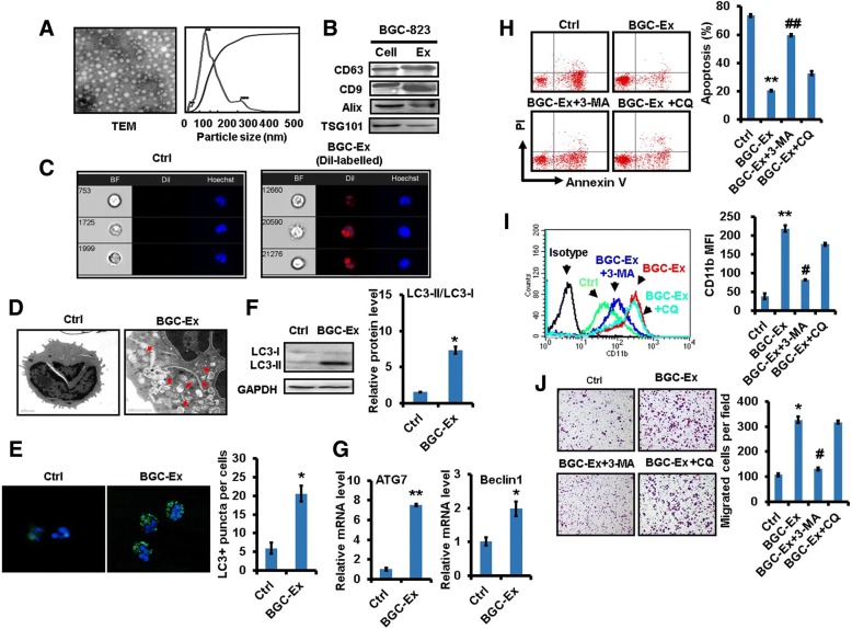
Background: Exosomes are extracellular vesicles that mediate cellular communication in health and diseases. Neutrophils could be polarized to a pro-tumor phenotype by tumor. The function of tumor-derived exosomes in neutrophil regulation remains unclear.

Methods: We investigated the effects of gastric cancer cell-derived exosomes (GC-Ex) on the pro-tumor activation of neutrophils and elucidated the underlying mechanisms.

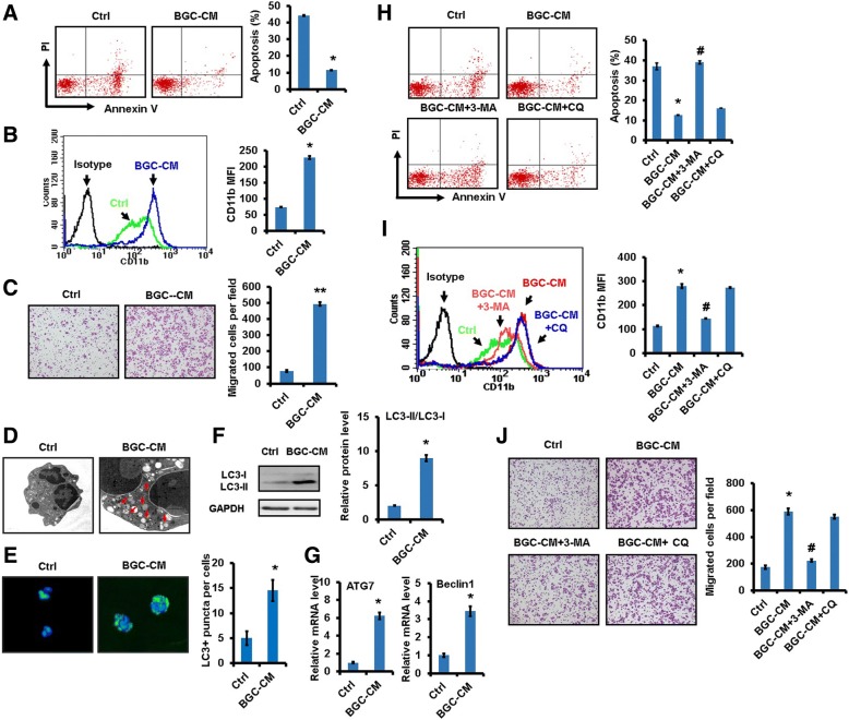
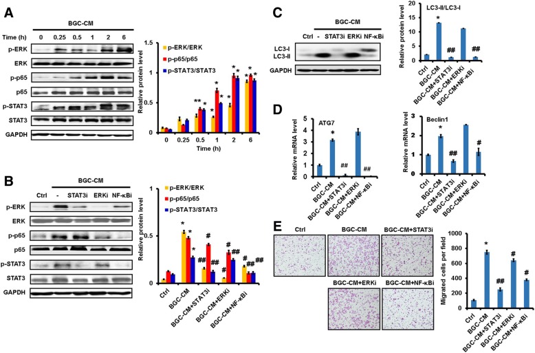
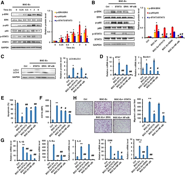
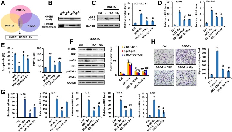
Results: GC-Ex prolonged neutrophil survival and induced expression of inflammatory factors in neutrophils. GC-Ex-activated neutrophils, in turn, promoted gastric cancer cell migration. GC-Ex transported high mobility group box-1 (HMGB1) that activated NF-κB pathway through interaction with TLR4, resulting in an increased autophagic response in neutrophils. Blocking HMGB1/TLR4 interaction, NF-κB pathway, and autophagy reversed GC-Ex-induced neutrophil activation. Silencing HMGB1 in gastric cancer cells confirmed HMGB1 as a key factor for GC-Ex-mediated neutrophil activation. Furthermore, HMGB1 expression was upregulated in gastric cancer tissues. Increased HMGB1 expression was associated with poor prognosis in patients with gastric cancer. Finally, gastric cancer tissue-derived exosomes acted similarly as exosomes derived from gastric cancer cell lines in neutrophil activation.

Conclusion: We demonstrate that gastric cancer cell-derived exosomes induce autophagy and pro-tumor activation of neutrophils via HMGB1/TLR4/NF-κB signaling, which provides new insights into mechanisms for neutrophil regulation in cancer and sheds lights on the multifaceted role of exosomes in reshaping tumor microenvironment.

Keywords: Activation; Autophagy; Exosome; Gastric cancer; Neutrophil; Pro-tumor.


Conflict of interest statement

Ethics approval and consent to participate

The use of clinical samples was approved by the ethics committee of Jiangsu University and informed consent was obtained from all patients.

Consent for publication

All of the authors are aware of and agree to the content of the paper and their being listed as a co-author of the paper.

Competing interests

The authors declare that they have no competing interests.

Publisher’s Note

Springer Nature remains neutral with regard to jurisdictional claims in published maps and institutional affiliations.

Figures

Fig. 1
Fig. 1
Gastric cancer cell-derived conditioned medium induced autophagy and pro-tumor activation of neutrophils. a. Flow cytometric analyses for apoptosis in neutrophils treated with or without conditioned medium from BGC-823 gastric cancer cells (BGC-CM). b. The expression of CD11b in BGC-CM-treated neutrophils was determined by flow cytometric analysis. c. Transwell migration assays for gastric cancer cells following treatment with supernatant from BGC-CM-treated neutrophils. d. Transmission electron microscopy analyses of autophagosomes (arrows) in neutrophils treated with BGC-CM. Scale bar=1 μm. e. Immunofluorescent staining of LC3-positive puncta in neutrophils treated with or without BGC-CM. Nuclei were counterstained by Hoechst 33342 (blue). f. Western blot assays for the expression of LC3-II in neutrophils incubated with conditioned medium from gastric cancer cells. g. The expression of ATG7 and BECN1 genes in neutrophils treated with conditioned medium from gastric cancer cells was determined by qRT-PCR. h. Neutrophils were pre-treated with autophagy inhibitors 3-MA or CQ followed by incubation with BGC-CM. The percentage of apoptotic neutrophils was determined by using flow cytometry. i. FACS analyses of CD11b expression in neutrophils treated with 3-MA and CQ prior to exposure to BGC-CM. j. Gastric cancer cells were incubated with the supernatants from BGC-CM-treated neutrophils that were pre-treated with or without autophagy inhibitors 3-MA or CQ and used for transwell migration assay. **P<0.01 and *P<0.05 compared to control (Ctrl); ##P<0.01 and #P<0.05 compared to BGC-CM.
Fig. 2
Fig. 2
Gastric cancer cell-derived conditioned medium induced pro-tumor activation of neutrophils through NF-κB pathway. a. Western blot assays for the expression of p-p65, p-STAT3, and p-ERK in neutrophils treated with GC-CM for different times. b-e. Neutrophils were pre-treated with inhibitors for NF-κB, STAT3, and ERK pathways followed by incubation with BGC-CM. The expression of phosphorylated NF-κB, STAT3, and ERK in neutrophils was determined by western blot (b). Western blot assays for LC3-II expression in neutrophils (c). QR-PCR analyses of ATG7 and BECN1 expression in neutrophils (d). Gastric cancer cells were incubated with the supernatants from neutrophils and used for transwell migration assay (e). **P<0.01 and *P<0.05 compared to control; ##P<0.01 and #P<0.05 compared to BGC-CM
Fig. 3
Fig. 3
Gastric cancer cell-derived exosomes induced autophagy to promote pro-tumor activation of neutrophils. a. The morphology (left) and size (right) of gastric cancer cell-derived exosomes were determined by TEM and NTA, respectively. b. The expression of exosomal markers CD9 and CD63 was determined by western blot. c. The internalization of CM-Dil-labelled exosomes (red) by neutrophils was determined by Imaging flow cytometry. Nuclei were counterstained with Hoechst 33342 (blue). BF, bright field. d. The presence of autophagosomes (arrows) in neutrophils treated with gastric cancer cell-derived exosomes (BGC-Ex) was determined by TEM. Scale bar=1 μm. e. Immunofluorescent staining of LC3-positive puncta (green) in BGC-Ex-treated neutrophils. f. The expression of LC3-II in BGC-Ex-treated neutrophils was confirmed by western blot. g. QRT-PCR analyses of ATG7 and BECN1 expression in BGC-Ex-treated neutrophils. h-j. Neutrophils were pre-treated with autophagy inhibitors 3-MA and CQ followed by treatment with BGC-Ex. FACS analyses of spontaneous apoptosis in neutrophil were shown (h). CD11b expression in neutrophils was determined by FACS analyses (i). Gastric cancer cells were incubated with the supernatants from neutrophils and used for transwell migration assay (j). **P<0.01 and *P<0.05 compared to control (Ctrl); ##P<0.01 and #P<0.05 compared to BGC-Ex
Fig. 4
Fig. 4
Gastric cancer cell-derived exosomes induced pro-tumor activation of neutrophils through NF-κB pathway. a. Western blot assays for the expression of p-p65, p-STAT3, and p-ERK in neutrophils treated with GC-Ex for different times. b-h. Neutrophils were pre-treated with inhibitors for NF-κB, STAT3, and ERK pathways followed by incubation with BGC-Ex. The expression of phosphorylated NF-κB, STAT3, and ERK in neutrophils was determined by western blot (b). Western blot assays for the expression of LC3-II in neutrophils (c). The expression of ATG7 and BECN1 genes in neutrophils was determined by qRT-PCR (d). Flow cytometric analyses for apoptosis in neutrophils (e). The expression of CD11b in neutrophils (f). QRT-PCR analyses of pro-inflammatory factor gene expression (IL-1β, IL-6, IL-8, OSM, and TNFα) in neutrophils (g).Transwell migration assays for gastric cancer cells following treatment with supernatant from neutrophils (h). **P<0.01 and *P<0.05 compared to control (Ctrl); ##P<0.01 and #P<0.05 compared to BGC-Ex
Fig. 5
Fig. 5
Gastric cancer cell-derived exosomes induced neutrophil activation through interaction of HMGB1 and TLR4. a. LC-MS/MS proteomic analyses for exosomes from gastric cancer cell lines (BGC-823, MGC80-3, and SGC-7901). b. The expression of HMGB1 in gastric cancer cell-derived exosomes was verified by western blot. c-h. Neutrophils were pre-treated with HMGB1 antagonist (Gly) or TLR4 inhibitor (TAK) followed by treatment with BGC-Ex. Western blot assays for the expression of LC3-II in neutrophils (c). The expression of ATG7 and BECN1 genes in neutrophils was determined by qRT-PCR (d). Flow cytometric analyses for apoptosis and CD11b expression in neutrophils (e). The expression of phosphorylated NF-κB, STAT3, and ERK in neutrophils was determined by western blot (f). QRT-PCR analyses of pro-inflammatory factor gene expression (IL-1β, IL-6, IL-8, OSM, and TNFα) in neutrophils (g).Transwell migration assays for gastric cancer cells following treatment with supernatant from neutrophils (h). **P<0.01 and *P<0.05 compared to control (Ctrl); ##P<0.01 and #P<0.05 compared to BGC-Ex
Fig. 6
Fig. 6
Gastric cancer tissue-derived exosomes induced pro-tumor activation of neutrophils. a. In silicon analyses of HMGB1 expression in paired gastric cancer and non-cancerous tissues from GEO dataset (GSE13911). b. The expression of HMGB1 in gastric cancer tissues and paired non-cancerous tissues was measured by ELISA (n=9). c. Tissue microarray analyses of HMGB1 expression in paired gastric cancer tissues and non-cancerous tissues (n=76). Survival time of gastric cancer patients was analyzed by Kaplan-Meier analysis. d-e. The percentage of apoptotic cells (d) and CD11b expression (e) in neutrophils treated with gastric cancer tissue-derived exosomes (T-Ex) and non-cancerous tissue-derived exosomes (N-Ex) were determined by flow cytometry. f. QRT-PCR analyses of ATG7 and BECN1 expression in neutrophils treated with T-Ex and N-Ex. g. Western blot assays for the activation of NF-κB, STAT3, and ERK pathways in neutrophils treated with T-Ex and N-Ex. h. Gastric cancer cells were incubated the supernatants from T-Ex- and N-Ex-treated neutrophils and used for transwell migration assay. i-k. Neutrophils were treated with T-Ex in the presence or absence of HMGB1 antagonist (Gly). The expression of ATG7 and BECN1 (i) and inflammatory factors (j) is measured by qRT-PCR. k. Transwell migration assays for gastric cancer cells following treatment with supernatant from neutrophils. *P<0.05 compared to control (Ctrl); ##P<0.01 and #P<0.05 compared to T- Ex
Fig. 7
Fig. 7
HMGB1 knockdown impaired neutrophil activation by gastric cancer cell-derived exosomes. HMGB1 was silenced in gastric cancer cell by siRNA. Neutrophils were treated with exosomes from HMGB1-silenced (si-HMGB1) and scramble control siRNA (si-Scr) transfected gastric cancer cells. a. Western blot assays for HMGB1 expression in gastric cancer cells transfected with HMGB1 siRNA and their derived exosomes. b-c. The percentage of apoptotic cells (b) and CD11b expression (c) in neutrophils were determined by FACS analyses. d. Western blot assays for LC3-II expression in neutrophils. e. The expression of ATG7 and BECN1 genes is measured by qRT-PCR. f. The expression of p-p65, p-STAT3 and p-ERK in neutrophils was determined by western blot. g. The expression of inflammatory factors in neutrophils was measured by qRT-PCR. h. Gastric cancer cells was incubated with the supernatants from neutrophils and subjected to transwell migration assay. i. Proposed model for the role of tumor-derived exosomes in the pro-tumor activation of neutrophils to promote gastric cancer cell migration. **P<0.01 and *P<0.05 compared to control (Ctrl); ##P<0.01 and #P<0.05 compared to si-Scr Ex

References

    1. Powell DR, Huttenlocher A. Neutrophils in the tumor microenvironment. Trends Immunol. 2016;37:41–52. doi: 10.1016/j.it.2015.11.008. - DOI - PMC - PubMed
    1. Liang W, Ferrara N. The complex role of neutrophils in tumor angiogenesis and metastasis. Cancer Immunol Res. 2016;4:83–91. doi: 10.1158/2326-6066.CIR-15-0313. - DOI - PubMed
    1. Swierczak A, Mouchemore KA, Hamilton JA, Anderson RL. Neutrophils: important contributors to tumor progression and metastasis. Cancer Metastasis Rev. 2015;34:735–751. doi: 10.1007/s10555-015-9594-9. - DOI - PubMed
    1. Houghton AM, Rzymkiewicz DM, Ji H, Gregory AD, Egea EE, Metz HE, et al. Neutrophil elastase-mediated degradation of IRS-1 accelerates lung tumor growth. Nat Med. 2010;16:219–223. doi: 10.1038/nm.2084. - DOI - PMC - PubMed
    1. Antonio N, Bonnelykke-Behrndtz ML, Ward LC, Collin J, Christensen IJ, Steiniche T, et al. The wound inflammatory response exacerbates growth of pre-neoplastic cells and progression to cancer. EMBO J. 2015;34:2219–2236. doi: 10.15252/embj.201490147. - DOI - PMC - PubMed

Publication types

MeSH terms