Prevalence of Pneumatized Articular Tubercle Using Panoramic Radiography and Cone Beam-Computed Tomography: A Retrospective Study
- PMID: 30294148
- PMCID: PMC6169291
- DOI: 10.4103/ccd.ccd_64_18
Prevalence of Pneumatized Articular Tubercle Using Panoramic Radiography and Cone Beam-Computed Tomography: A Retrospective Study
Abstract
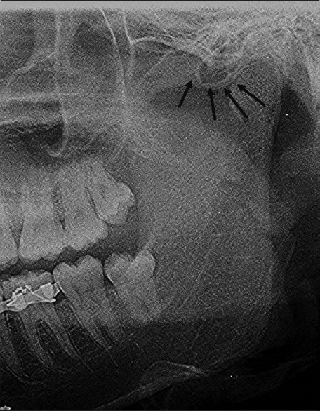
Context: In 1985, Tyndall and Matteson first described the air cells that occur in the root of zygomatic arch and in the articular eminence of the temporal bone but do not extend beyond the zygomaticotemporal suture.
Aims: The aim is to study the prevalence and patterns of pneumatized articular tubercle (PAT) retrospectively using two different imaging modalities, i.e., digital panoramic radiography and cone-beam computed tomography (CBCT).
Subjects and methods: Total 3000 panoramic radiographs belonging to 1291 females and 1709 males and CBCT scans of 200 patients belonging to 62 females and 138 males were studied retrospectively and investigated for radiographic features of pneumatized articular tubercle.
Results: Prevalence found by using panoramic radiography is 1.96% and by using CBCT is 12.5%.
Conclusions: This research scrutinizes the prevalence of pneumatized articular tubercle and establishes the prevalence of the same among the studied population, using panoramic radiography and CBCT.
Keywords: Articular tubercle; cone-beam computed tomography; panoramic radiography; pneumatization.
Conflict of interest statement
There are no conflicts of interest.
Figures


Similar articles
-
Pneumatization of the articular eminence in cone-beam computed tomography: prevalence and characteristics - literature review.Folia Morphol (Warsz). 2023;82(2):242-247. doi: 10.5603/FM.a2022.0023. Epub 2022 Mar 14. Folia Morphol (Warsz). 2023. PMID: 35285512
-
Accuracy of digital panoramic radiography in the diagnosis of temporal bone pneumatization: a study in vivo using cone-beam-computed tomography.J Craniomaxillofac Surg. 2014 Jul;42(5):477-81. doi: 10.1016/j.jcms.2013.06.005. Epub 2013 Jul 11. J Craniomaxillofac Surg. 2014. PMID: 23850158
-
Pneumatized Articular Eminence and Assessment of Its Prevalence and Features on Panoramic Radiographs.J Dent (Tehran). 2015 Apr;12(4):235-42. J Dent (Tehran). 2015. PMID: 26622277 Free PMC article.
-
Pneumatization of the articular eminence on cone beam computed tomography: prevalence, characteristics and a review of the literature.Dentomaxillofac Radiol. 2011 Feb;40(2):110-4. doi: 10.1259/dmfr/75842018. Dentomaxillofac Radiol. 2011. PMID: 21239574 Free PMC article. Review.
-
[Use of diagnostic modalities for dentofacial imaging in forensic dentistry. Literature review].Rev Cient Odontol (Lima). 2021 Dec 9;9(4):e088. doi: 10.21142/2523-2754-0904-2021-088. eCollection 2021 Oct-Dec. Rev Cient Odontol (Lima). 2021. PMID: 38463727 Free PMC article. Review. Spanish.
Cited by
-
Comparison of mastoid air cell volume in patients with or without a pneumatized articular tubercle.Imaging Sci Dent. 2022 Mar;52(1):27-32. doi: 10.5624/isd.20210153. Epub 2021 Nov 18. Imaging Sci Dent. 2022. PMID: 35387098 Free PMC article.
-
Prevalence of pneumatization of the articular eminence and glenoid fossa viewed on cone-beam computed tomography examinations in a Turkish sample.Oral Radiol. 2020 Jan;36(1):40-46. doi: 10.1007/s11282-019-00378-1. Epub 2019 Feb 22. Oral Radiol. 2020. PMID: 30796675
References
-
- Tyndall DA, Matteson SR. The zygomatic air cell defect (ZACD) on panoramic radiographs. Oral Surg Oral Med Oral Pathol. 1987;64:373–6. - PubMed
-
- Tremble EG. Pneumatization of the temporal bone. Arch Otolaryngol. 1934;19:172–82.
-
- Hollinshead WH. Anatomy for Surgeons. 2nd ed. Vol. 1. New York: Harper SC Row, Publishers; 1968. The Head and Neck; pp. 190–4.
-
- Patil K, Mahima VG, Malleshi SN, Srikanth HS. Prevalence of zygomatic air cell defect in adults – A retrospective panoramic radiographic analysis. Eur J Radiol. 2012;81:957–9. - PubMed
-
- Abramovitch K, Rice DD. Basic principles of cone beam computed tomography. Dent Clin North Am. 2014;58:463–84. - PubMed