Rhinoscleroma: Report of an Erratic Palatal Swelling
- PMID: 30294174
- PMCID: PMC6169265
- DOI: 10.4103/ccd.ccd_282_18
Rhinoscleroma: Report of an Erratic Palatal Swelling
Abstract
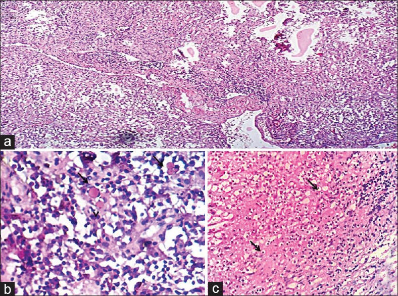
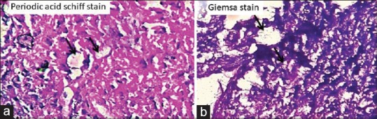
Clinicians come across cases of palatal swellings that present with ambiguous features. They may vary in their etiology from numerous infectious and noninfectious causes to a wide array of neoplasms. Accurate diagnosis of such lesions is vital for their prompt and precise management. Rhinoscleroma (RS), as its name suggests, is a persistent, specific, granulomatous disease that results in sclerosis of the affected organ - most frequently the nose. Although its occurrence in the adjacent sites has been reported, the clinical findings did not offer much deviation from the expected. Reported here is a case of RS involving the palate which not only detoured from its usual site and course of spread but also gave off a confusing façade in terms of its clinical presentation.
Keywords: Chronic granulomatous inflammation; palatal swellings; rhinoscleroma.
Conflict of interest statement
There are no conflicts of interest.
Figures


Similar articles
-
Rhinoscleroma: a detailed histopathological diagnostic insight.Int J Clin Exp Pathol. 2015 Jul 1;8(7):8438-45. eCollection 2015. Int J Clin Exp Pathol. 2015. PMID: 26339415 Free PMC article.
-
Palatal presentation of scleroma.J Laryngol Otol. 2001 Aug;115(8):679-80. doi: 10.1258/0022215011908621. J Laryngol Otol. 2001. PMID: 11535159 Review.
-
Rhinoscleroma--a diagnostic challenge.Laryngoscope. 1983 Jul;93(7):866-70. doi: 10.1288/00005537-198307000-00003. Laryngoscope. 1983. PMID: 6865620
-
[Apropos of a case of rhinoscleroma].Med Trop (Mars). 1984 Apr-Jun;44(2):165-9. Med Trop (Mars). 1984. PMID: 6090860 French.
-
Rhinoscleroma.Arch Pathol Lab Med. 2018 Dec;142(12):1533-1536. doi: 10.5858/arpa.2018-0073-RA. Epub 2018 Aug 31. Arch Pathol Lab Med. 2018. PMID: 30168726 Review.
References
-
- Rajendran R. Shafer's textbook of oral pathology. Elsevier India. 2009
-
- Bhagyalakshmi A, Rao CV, Krishna Kishore T, Kartheek BV. Rhinoscleroma: A case report. Int J Case Rep Images. 2014;5:868–72.
-
- Nayak P, Pramod RC, Suresh KV, Desai D, Pandit S, Ingaleshwar PS, et al. Rhinoscleroma of nose extruding into oral cavity. J Coll Physicians Surg Pak. 2015;25(Suppl 1):S27–9. - PubMed
-
- Basavannaiah S. Unusual presentation of rhinoscleroma mimicking midline granuloma: A histological surprise. Int Res J Clin Med. 2016;1:13–7.
-
- Efared B, Hammas N, Gabrielle AE, Ben Mansour N, El Fatemi H, Chbani L, et al. Rhinoscleroma: A chronic infectious disease of poor areas with characteristic histological features – Report of a series of six cases. Trop Doct. 2018;48:33–5. - PubMed