

Gadolinium Deposition in Brain: Current Scientific Evidence and Future Perspectives
- PMID: 30294259
- PMCID: PMC6158336
- DOI: 10.3389/fnmol.2018.00335
Gadolinium Deposition in Brain: Current Scientific Evidence and Future Perspectives
Abstract
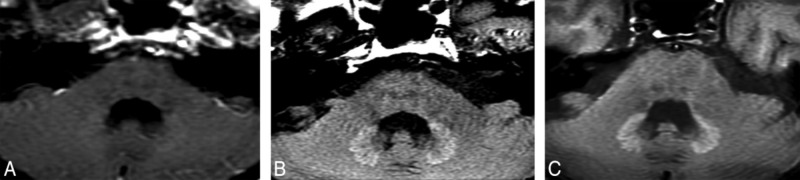
In the past 4 years, many publications described a concentration-dependent deposition of gadolinium in the brain both in adults and children, seen as high signal intensities in the globus pallidus and dentate nucleus on unenhanced T1-weighted images. Postmortem human or animal studies have validated gadolinium deposition in these T1-hyperintensity areas, raising new concerns on the safety of gadolinium-based contrast agents (GBCAs). Residual gadolinium is deposited not only in brain, but also in extracranial tissues such as liver, skin, and bone. This review summarizes the current evidence on gadolinium deposition in the human and animal bodies, evaluates the effects of different types of GBCAs on the gadolinium deposition, introduces the possible entrance or clearance mechanism of the gadolinium and potential side effects that may be related to the gadolinium deposition on human or animals, and puts forward some suggestions for further research.
Keywords: T1 hyperintensity; brain; gadolinium deposition; gadolinium-based contrast agents; magnetic resonance imaging.
Figures
References
-
- Bjørnerud A., Vatnehol S., Larsson C., Duetønnessen P., Hol P. K., Groote I. R. (2017). Signal enhancement of the dentate nucleus at unenhanced MR imaging after very high cumulative doses of the macrocyclic gadolinium-based contrast agent gadobutrol: an observational study. Radiology 285 434–444. 10.1148/radiol.2017170391 - DOI - PubMed
Publication types
LinkOut - more resources
Full Text Sources
Other Literature Sources
Miscellaneous